Introduction
Escherichia coli (E coli) is a species of gram-negative, rod-shaped bacteria belonging to the genus Escherichia and commonly residing in the colon of humans and many other animal species. Shigatoxigenic E coli (STEC) and verotoxigenic E coli (VTEC) are E coli strains known to produce Shiga toxin and Shiga-like toxin (verotoxin), respectively. The E coli strains that cause bloody diarrhea in humans are collectively known as enterohemorrhagic E coli (EHEC).[1] These three terms are often used interchangeably. These pathogens are clinically significant due to their potential to cause diarrhea, hemorrhagic colitis, hemolytic uremic syndrome (HUS) and thrombotic thrombocytopenia purpura (TTP), and contribute to outbreaks via foodborne, waterborne, animal-to-human, and person-to-person transmission.[2][3][4] EHEC serotype O157:H7 is the most common cause of HUS in the United States, but other EHEC strains, including E coli 026, can cause HUS.[5]
Etiology
Register For Free And Read The Full Article
 Search engine and full access to all medical articles
- 10 free questions in your specialty
- Free CME/CE Activities
- Free daily question in your email
- Save favorite articles to your dashboard
- Emails offering discounts
Learn more about a Subscription to StatPearls Point-of-Care
Etiology
E coli is a gram-negative, rod-shaped bacterium belonging to the genus Escherichia. This organism contains up to 2,000 genes that encode various virulence factors, reflecting the diversity of E coli clones, including EHEC.[6] E coli is a facultative anaerobe, measuring 1 to 2 μm in length and 0.5 μm in width, with chemotactic motility. This microorganism commonly colonizes the intestines of all known mammals.[7][8][9] For further information on Escherichia coli, refer to StatPearls' companion topic, "Escherichia coli infection."
E coli is one of the most commonly identified bacteria in the human intestinal microbiota from birth and remains a lifelong colonizer.[10] These strains are likely transmitted to humans from the gut colonization of ruminants, particularly farm animals.[11] Humans can become infected through environmental transmission from contaminated food and water or close contact with infected animals or individuals. The contamination of fresh fruits and vegetables occurs secondary to fecal contamination in agricultural irrigation water or runoff. E coli O157:H7 has hardy survival characteristics exceeding those found in commensal E coli strains, which enable this food-borne pathogen to survive a wide range of harsh conditions frequently encountered within the human food chain. This pathogen can persist for extended periods in the food matrix.[12]
Epidemiology
The Centers for Disease Control and Prevention (CDC) estimates that EHEC was responsible for over 350,000 illnesses in 2019 (90% credible interval, 159,000 - 648,000), of which approximately one-quarter were caused by the O157 strain and three-quarters due to non-O157 strains.[13] Over a 20-year period, E coli O157:H7 outbreaks in the U.S. resulted in 17% of the cohort becoming hospitalized, with 4% resulting in HUS.[14] The 2022 preliminary report from the 10 U.S. sites of the Foodborne Diseases Active Surveillance Network (FoodNet) reported an annual incidence of 5.7 cases of EHEC per 100,000 people in the U.S., increased from the average annual incidence of 5.3 cases per 100,000 people from 2016-2018.[15] Children younger than 5 years have the highest incidence of EHEC infection as well as the highest risk of subsequent HUS.
A 2014 review of studies from 10 out of 14 World Health Organization sub-regions estimated that EHEC causes a global incidence of 2.8 million cases per year, leading to nearly 4000 cases of HUS and 230 deaths per year.[16] The economic burden of illness caused by this bacterium, resulting from medical expenses, mortality, and lost productivity, is estimated to be $405 million per year.[17]
The intestines of ruminants are the natural reservoir for E coli O157:H7, and outbreaks can occur from ingesting undercooked meat or fomites from manure-contaminated food or water. Contamination can also result from the use of manure as fertilizer or from water supplies contaminated by runoff from cattle farms. Although variation in fecal shedding of E coli O157:H7 has been reported, ranging from 0% to 80% among the cattle population, a seasonal pattern has been observed, with prevalence increasing during the summer months.[18]
Pathophysiology
Upon entry, EHEC migrates to the gastrointestinal tract, where it survives innate host defenses, including saliva, gastric acids, and intestinal mucus, by utilizing acid resistance mechanisms.[19] The organism targets the Peyer patches and intestinal villi, where it forms pathogenic lesions and colonizes the large intestine.[20] In this environment, virulence factors are upregulated through interactions with short-chain fatty acids secreted by intestinal flora, facilitating further adherence and increasing toxin susceptibility.[21][22]
EHEC strains produce Shiga-like toxins, which disrupt membrane ion channels in the epithelial membrane of the intestine. This dysregulation leads to ion loss and a massive loss of water, potentially allowing for bacterial translocation and invasion.[23] The toxin also functions as a cell transducer and immune modulator, inducing pro-inflammatory and proapoptotic sequelae. Additionally, this toxin can inactivate 60S ribosomal units, inhibiting protein synthesis in endothelial cells.[24]
Neutrophil numbers rise markedly, and the extent of this increase correlates with a higher occurrence of HUS.[25] Inflammatory monocytes also rise and produce pro-inflammatory cytokines. Shiga toxin-susceptible receptors are present on erythrocytes, platelets, and monocytes.[26][27] Microthrombi may develop due to the interaction between Shiga toxin and platelet-leukocyte aggregation. Consequently, activated endothelial cells may become thrombogenic, leading to endothelial lesions in the microvasculature, primarily in the kidneys, and less frequently in other organs, contributing to the development of HUS. Thrombocytopenia, a characteristic feature of HUS pathogenesis, may be linked to the consumption of microthrombi by the immune response. In severe cases, nonimmune microangiopathic hemolytic anemia (MAHA) may occur.[28]
Endothelial dysfunction in the kidneys can result in acute renal impairment. Although the kidney and gastrointestinal tract are the most commonly affected organs in HUS, studies have also shown evidence of involvement in the central nervous system, pancreas, skeletal system, and myocardium. While the mechanism of microvascular injury is not fully understood, evidence suggests that verocytotoxin plays a role in mediating cell injury, altering the endothelial cell's normal anticoagulant profile to a procoagulant state.[29]
After an E coli infection, several factors determine the progression of the disease to HUS, including the following:
- Bacterial strain: Serotype O157:H7 is most often responsible for the progression to HUS.
- Age: The rate of progression to HUS is higher in young children. A study found that the progression rate was 12.9% in children under 5 years, 6.8% in children aged 5 to 10 years, and 8% in children older than 10 years.[30]
- Antibiotic therapy: Treatment of E coli O157:H7 with antibiotics, particularly β-lactams, may increase the risk of developing HUS.[31]
- Environmental factors: Variables, such as proximity to cattle density and rainfall, have been identified in observational datasets. However, such factors should be considered in the context of E coli transmission.[32][33]
- Genetic factors: The presence of a platelet glycoprotein 1b α 145M allele has been associated with an increased risk of HUS.[34]
Other factors that may correlate with HUS include a higher leukocyte count and vomiting during the first week of illness.[35] For further information on E coli pathophysiology, refer to StatPearls' companion topic, "Escherichia coli infection."
Histopathology
In the acute phase of HUS, kidney specimens show microvascular injury, characterized by microthrombi deposition and detached, swollen glomerular endothelial cells associated with inflammatory cell infiltration. Similar changes have been observed in other organs, including the pancreas, adrenal glands, and brain.[36] Autopsy findings have included platelet aggregation, fibrin accumulation, and a low platelet count on factor VIII staining. Areas of ischemia with microscopic angiopathy may be present, and destruction of the renal cortex can occur, showing capillary wall thickening, thrombosis of the capillary lumen, preglomerular arteries, and endothelial cells.[37] Gastrointestinal changes may include mucosal and submucosal edema or hemorrhage.[38]
Toxicokinetics
EHEC is not invasive, making bacteremia rare. This microorganism adheres to mammalian cells and secretes bacterial proteins into host cells via a type III secretion system. Shiga-like toxins 1 and 2 are secreted and are responsible for organ damage. Shiga-like toxin 2 is more frequently associated with severe disease.[39]
Shiga toxin consists of 2 subunits, A and B. Proteolysis degrades subunit A into A1 and A2. In target organs, such as the kidney, brain, and gut, subunit B attaches to glycolipid receptors on the cell surface. In humans, these receptors are identified as Gb3, primarily expressed in kidney tubular cells, the brain, and gut epithelium. Tumor necrosis factor alpha amplifies the cytotoxicity in the kidney.[40]
After binding to the cell surface, Shiga toxin is endocytosed and transported in a retrograde direction to the Golgi apparatus and endoplasmic reticulum. From there, the toxin is translocated to the cytosol, where it inactivates ribosomes, leading to cell death, sloughing of the mucosa, and eventual bloody diarrhea.[41] Other virulence factors encoded on plasmids include enterohemolysin, which has a cytolytic effect; extracellular serine protease, which cleaves human coagulation factor V; and enteroaggregative E coli heat-stable enterotoxin 1, which may contribute to the development of STEC diarrhea.[42][43]
History and Physical
A history of exposure to contaminated sources, including food and drinking water, or close contact with ruminants, is often reported. EHEC clinically manifests as bloody or watery diarrhea without fever and typically a white blood cell count above 10,000/μL, sometimes associated with abdominal cramping. Diarrhea initially may not be bloody, often being watery in consistency. Most patients will not manifest a temperature during the initial presentation and evaluation. As a result of nausea, vomiting, and profuse diarrhea, patients will often note dehydration, asthenia, and decreased urine output. Systemic signs of dehydration such as dry mucous membranes, tachycardia, decreased skin turgor, slow capillary refill, cold extremities, and delirium, presage worsened morbidity, particularly in children. The incubation period between exposure to EHEC and the onset of symptoms is typically 3 to 4 days.[44]
HUS is a major complication of EHEC infection, characterized by the clinical triad of anemia due to hemolysis, impaired renal function, and thrombocytopenia, primarily affecting young children. Anemia typically manifests as pallor on examination, thrombocytopenia as petechial rashes, and a decline in renal function as decreased urine output. However, atypical cases may not present with all these features and may also warrant consideration of alternative diagnoses.[45] HUS following bloody diarrhea secondary to EHEC is called "D+ HUS" or "typical HUS," while HUS caused by other factors is termed "D- HUS" or "atypical hemolytic uremic syndrome" (aHUS).[46]
Evaluation
Initial laboratory evaluation should include a complete blood count to rule out leukocytosis, hemolysis, and thrombocytopenia. A complete metabolic profile will aid in ruling out dehydration, electrolyte disturbance, and uremia. The majority of patients with E coli 0157:H7 colitis will have a leukocytosis above 10,000/microL.
Patients suspected of EHEC infection should be tested for Shiga toxin or EHEC through stool culture within the first days after onset. Culture the diarrheal specimen with sorbitol-MacConkey agar or multiindicator chromogenic agar. Shiga toxin is primarily detected using a direct enzyme immunoassay; however, the genes encoding this toxin can also be identified by real-time PCR.[47] Some centers use matrix-assisted laser-desorption/ionization time-of-flight mass spectrometry (MALDI-TOF MS) to detect the genes.
Due to the risk of Coombs-negative MAHA, a hemolysis screen is warranted in cases of anemia. A blood film may reveal red cell fragmentation, and hypocomplementemia may occur. ADAMST13 does not typically decrease in EHEC HUS, and a reduction in ADAMST13 is more commonly associated with aHUS.[48]
Treatment / Management
Supportive treatment is essential for patients with EHEC diarrhea. Replacing electrolytes and water is particularly important for those with D+ HUS, which may be achieved through oral or intravenous fluid and electrolyte administration. Most enterohemorrhagic E coli diarrheal patients recover within ten days without treatment other than fluid replacement. Early intervention with close and judicious monitoring of volume and sodium status can help reduce the risk of progressing to oliguric or anuric HUS.[49]
Antibiotics, particularly β-lactams, are relatively contraindicated in EHEC-associated HUS, as they may indirectly cause the release of Shiga toxin from lysed bacteria, resulting in further renal and gastrointestinal injury.[50] The use of β-lactam antibiotics has also been linked to the development of HUS.
Antiperistaltic agents, such as loperamide or dicyclomine, slow intestinal motility and increase the risk of systemic complications; clinicians should avoid their utilization in this setting.[51]
Medications that may exacerbate renal impairment, including antihypertensives, should be withheld during this period, as they can impair renal perfusion.[52] Advancements in dialysis and intensive care have significantly reduced mortality, especially among young children. Up to 2/3 of children infected with EHEC may require dialysis.[53] Peritoneal dialysis is often the best option for children with acute and severe renal impairment and significant bloody diarrhea.
Bilateral nephrectomy may be life-saving in severe cases where the kidneys are the primary site of disease involvement. This intervention can help control the spread of microvascular lesions, particularly in therapy-resistant malignant hypertension.
Given the often severe prognosis, immediate supportive treatment is crucial to improve outcomes. Additional supportive treatments for HUS depend on the patient’s symptoms and may include the following:
- Red blood cell transfusions, particularly in those with anemia
- Plasma exchange [54]
- Fresh frozen plasma
- Eculizumab, particularly in those with neurological manifestations [55]
The effect of plasma exchange is most notable in older adults and children when initiated early in the disease course.[56] Fresh frozen plasma has been employed in rare cases.[57] Eculizumab has also been used for typical HUS with neurological involvement.(B3)
Platelet transfusions are generally contraindicated due to the risk of exacerbating illness. Transfusions may perpetuate platelet aggregation in patients with thrombotic microangiopathy associated with HUS.[58][59]
Differential Diagnosis
In patients of unusual age or without a history of diarrhea, anomalous or atypical E coli HUS should be considered. Acute bloody diarrhea may also suggest other differentials, including inflammatory bowel disease, rectal or colorectal carcinoma, hemorrhoids, and a perforated viscus. Bloody diarrhea can also result from infections caused by other organisms, including Salmonella, Campylobacter, Yersinia, tuberculosis, and Entamoeba.[60] Noninfectious etiologies of hemorrhagic diarrhea, such as ischemic colitis, mesenteric ischemia, Crohn disease, and ulcerative colitis, merit consideration as well.
Prognosis
Enterohemorrhagic E coli colitis has a good prognosis for recovery when patients do not have systemic manifestations of diarrheal illness. Early diagnosis of EHEC infection and prompt fluid replacement have been shown to improve long-term outcomes by reducing kidney damage. The volume of appropriate intravenous fluid replacement is directly associated with the risk of developing oliguria and anuria in patients with EHEC-associated HUS.
Patients infected with the E coli O157:H7 serotype are more likely to present with hematochezia and leukocytosis than individuals unaffected by this strain. These patients also tend to require a longer duration of dialysis. Advancements in dialysis therapy and improved interventions for critically ill children have significantly reduced the acute mortality of HUS. However, as survival rates improve, chronic complications in long-term survivors are becoming increasingly apparent.[61]
The mortality rate for postdiarrheal HUS is approximately 3 to 5%.[62][63] Risk factors for mortality include high leukocyte count, high hematocrit, recent respiratory tract infection, hyponatremia, and oliguria.[64][65]
Complications
EHEC-associated bloody diarrhea often resolves without long-term consequences. However, the prognosis is severe in patients who develop HUS. Following treatment for HUS, some children may experience permanent loss of renal function, necessitating long-term renal replacement therapies. Even patients who recover baseline renal function remain at risk for the late onset of renal disease. Residual extrarenal complications may occur in some children, including neurological defects, insulin-dependent diabetes mellitus, pancreatic insufficiency, and gastrointestinal problems.[66] HUS is thus associated with significant mortality and multisystem morbidity. Attention should be given to extrarenal manifestations during the acute phase, and renal function should be closely monitored during the long-term follow-up of patients with HUS.[67]
Consultations
Nephrology consultation has merit if patients develop HUS, as up to 50% require hemodialysis if acute renal impairment occurs. Gastroenterology or infectious disease consultations may also provide expert guidance, especially in the initial diagnostic evaluation and patient care phase, when trying to differentiate EHEC from other infectious, inflammatory, or ischemic etiologies of bloody diarrhea.
Deterrence and Patient Education
Hand hygiene is one of the most important methods to prevent transmission of E coli and other causes of infectious diarrhea. All patients and caregivers should be counseled to perform regular hand hygiene after using the toilet or changing diapers, before and after preparing food, before eating, after handling garbage or other soiled items, and after touching animals, particularly in petting zoos. Healthcare workers tending to people with diarrhea should wear gowns and gloves in addition to stringent hand hygiene.
Implementing measures such as using drinkable water for food preparation, maintaining improved hygienic conditions during animal slaughter, adopting appropriate food processing techniques, properly cooking food, and educating food handlers and farm workers on food hygiene principles can significantly reduce the incidence of EHEC infections. Preventing foodborne diseases generally relies on good hygienic practices and controlling food contamination by biological and chemical hazards.
Preventing the spread of E coli 0157:H7 hemorrhagic colitis includes isolation of potentially infectious contacts in school or within institutions to minimize infectious transmission. Patients with diarrhea should be counseled to avoid swimming, food handling, and sexual activities and to practice strict hand hygiene.[68] In healthcare settings, these patients should be placed on contact precautions.[69]
Developing a human vaccine to prevent enterohemorrhagic E coli infection may eventually provide herd immunity, protect against HUS, and provide value in low-income, high-risk dysentery settings.
Pearls and Other Issues
EHEC is a foodborne disease that may be mitigated by practicing good hygiene and controlling food contamination. Public health and food standards authorities play a crucial role in regulating and monitoring safety related to foodborne contamination. In some jurisdictions, EHEC constitutes a public health notifiable condition. This human pathogen has been identified as a cause of bloody diarrhea outbreaks and HUS globally. Specific treatment options are unavailable, and therapeutic measures remain supportive.
Enhancing Healthcare Team Outcomes
The management of EHEC requires an interprofessional team approach, including an emergency department physician, an infectious disease consultant, a nephrologist, and an internist. Close fluid and electrolyte monitoring, facilitated by attentive nursing and medical care, is crucial for the early detection of clinical deterioration. Supportive treatment is sufficient for most patients, with particular attention to replacing electrolytes and water deficiencies, especially in those with D+ HUS.
Advancements in dialysis and intensive care have significantly reduced mortality, particularly in young children, where peritoneal dialysis may be necessary to manage severe complications. Surgical intervention, including bilateral nephrectomy, may be life-saving in severe cases. This procedure can help control the spread of microvascular lesions when the kidneys are the primary site of disease involvement, particularly in therapy-resistant malignant hypertension.
Given the potential severity of the prognosis, immediate supportive treatment may improve outcomes. Additional supportive therapies for patients with HUS are largely symptom-dependent and may include blood transfusions and, in rare cases, plasma exchange.
The relevant public health agencies should be notified of cases to facilitate contact tracing and environmental investigations. Collaboration with local government and food safety authorities is essential to identify and mitigate sources of exposure.
Media
(Click Image to Enlarge)
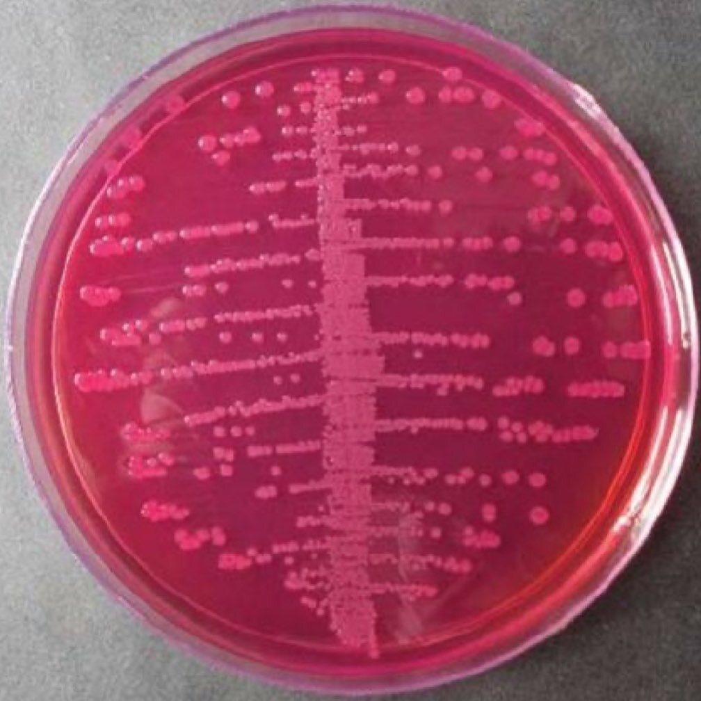
Escherichia coli on MacConkey agar. "Contributed by Muhammad Zubair, MBBS, FCPS"
References
Yang SC, Lin CH, Aljuffali IA, Fang JY. Current pathogenic Escherichia coli foodborne outbreak cases and therapy development. Archives of microbiology. 2017 Aug:199(6):811-825. doi: 10.1007/s00203-017-1393-y. Epub 2017 Jun 9 [PubMed PMID: 28597303]
Level 3 (low-level) evidenceHonish L, Punja N, Nunn S, Nelson D, Hislop N, Gosselin G, Stashko N, Dittrich D. Escherichia coli O157:H7 Infections Associated with Contaminated Pork Products - Alberta, Canada, July-October 2014. MMWR. Morbidity and mortality weekly report. 2017 Jan 6:65(52):1477-1481. doi: 10.15585/mmwr.mm6552a5. Epub 2017 Jan 6 [PubMed PMID: 28056011]
Crump JA, Sulka AC, Langer AJ, Schaben C, Crielly AS, Gage R, Baysinger M, Moll M, Withers G, Toney DM, Hunter SB, Hoekstra RM, Wong SK, Griffin PM, Van Gilder TJ. An outbreak of Escherichia coli O157:H7 infections among visitors to a dairy farm. The New England journal of medicine. 2002 Aug 22:347(8):555-60 [PubMed PMID: 12192014]
Luini MV, Colombo R, Dodaro A, Vignati C, Masia C, Arghittu M, Daprai L, Maisano AM, Vezzoli F, Bianchini V, Spelta C, Castiglioni B, Bertasi B, Ardissino G. Family Clusters of Shiga Toxin-producing Escherichia coli Infection: An Overlooked Source of Transmission. Data From the ItalKid-Hus Network. The Pediatric infectious disease journal. 2021 Jan:40(1):1-5. doi: 10.1097/INF.0000000000002877. Epub [PubMed PMID: 32898091]
Freedman SB, van de Kar NCAJ, Tarr PI. Shiga Toxin-Producing Escherichia coli and the Hemolytic-Uremic Syndrome. The New England journal of medicine. 2023 Oct 12:389(15):1402-1414. doi: 10.1056/NEJMra2108739. Epub [PubMed PMID: 37819955]
Kaper JB, Nataro JP, Mobley HL. Pathogenic Escherichia coli. Nature reviews. Microbiology. 2004 Feb:2(2):123-40 [PubMed PMID: 15040260]
Conway T, Cohen PS. Commensal and Pathogenic Escherichia coli Metabolism in the Gut. Microbiology spectrum. 2015 Jun:3(3):. doi: 10.1128/microbiolspec.MBP-0006-2014. Epub [PubMed PMID: 26185077]
Mittal N, Budrene EO, Brenner MP, Van Oudenaarden A. Motility of Escherichia coli cells in clusters formed by chemotactic aggregation. Proceedings of the National Academy of Sciences of the United States of America. 2003 Nov 11:100(23):13259-63 [PubMed PMID: 14597724]
Shiomi D, Mori H, Niki H. Genetic mechanism regulating bacterial cell shape and metabolism. Communicative & integrative biology. 2009 May:2(3):219-20 [PubMed PMID: 19641734]
Palmer C, Bik EM, DiGiulio DB, Relman DA, Brown PO. Development of the human infant intestinal microbiota. PLoS biology. 2007 Jul:5(7):e177 [PubMed PMID: 17594176]
Blanco JE, Blanco M, Blanco J. [Enterotoxigenic, verotoxigenic, and necrotoxigenic Escherichia coli in food and clinical samples. Role of animals as reservoirs of strains pathogenic for humans]. Microbiologia (Madrid, Spain). 1995 Mar:11(1):97-110 [PubMed PMID: 7546450]
Level 3 (low-level) evidenceThomas DE, Elliott EJ. Interventions for preventing diarrhea-associated hemolytic uremic syndrome: systematic review. BMC public health. 2013 Sep 3:13():799. doi: 10.1186/1471-2458-13-799. Epub 2013 Sep 3 [PubMed PMID: 24007265]
Level 3 (low-level) evidenceScallan Walter EJ, Cui Z, Tierney R, Griffin PM, Hoekstra RM, Payne DC, Rose EB, Devine C, Namwase AS, Mirza SA, Kambhampati AK, Straily A, Bruce BB. Foodborne Illness Acquired in the United States-Major Pathogens, 2019. Emerging infectious diseases. 2025 Apr:31(4):669-677. doi: 10.3201/eid3104.240913. Epub [PubMed PMID: 40133035]
Rangel JM, Sparling PH, Crowe C, Griffin PM, Swerdlow DL. Epidemiology of Escherichia coli O157:H7 outbreaks, United States, 1982-2002. Emerging infectious diseases. 2005 Apr:11(4):603-9 [PubMed PMID: 15829201]
Delahoy MJ, Shah HJ, Weller DL, Ray LC, Smith K, McGuire S, Trevejo RT, Scallan Walter E, Wymore K, Rissman T, McMillian M, Lathrop S, LaClair B, Boyle MM, Harris S, Zablotsky-Kufel J, Houck K, Devine CJ, Lau CE, Tauxe RV, Bruce BB, Griffin PM, Payne DC. Preliminary Incidence and Trends of Infections Caused by Pathogens Transmitted Commonly Through Food - Foodborne Diseases Active Surveillance Network, 10 U.S. Sites, 2022. MMWR. Morbidity and mortality weekly report. 2023 Jun 30:72(26):701-706. doi: 10.15585/mmwr.mm7226a1. Epub 2023 Jun 30 [PubMed PMID: 37384552]
Majowicz SE, Scallan E, Jones-Bitton A, Sargeant JM, Stapleton J, Angulo FJ, Yeung DH, Kirk MD. Global incidence of human Shiga toxin-producing Escherichia coli infections and deaths: a systematic review and knowledge synthesis. Foodborne pathogens and disease. 2014 Jun:11(6):447-55. doi: 10.1089/fpd.2013.1704. Epub 2014 Apr 21 [PubMed PMID: 24750096]
Level 1 (high-level) evidenceMarder Mph EP, Griffin PM, Cieslak PR, Dunn J, Hurd S, Jervis R, Lathrop S, Muse A, Ryan P, Smith K, Tobin-D'Angelo M, Vugia DJ, Holt KG, Wolpert BJ, Tauxe R, Geissler AL. Preliminary Incidence and Trends of Infections with Pathogens Transmitted Commonly Through Food - Foodborne Diseases Active Surveillance Network, 10 U.S. Sites, 2006-2017. MMWR. Morbidity and mortality weekly report. 2018 Mar 23:67(11):324-328. doi: 10.15585/mmwr.mm6711a3. Epub 2018 Mar 23 [PubMed PMID: 29565841]
Beauvais W, Gart EV, Bean M, Blanco A, Wilsey J, McWhinney K, Bryan L, Krath M, Yang CY, Manriquez Alvarez D, Paudyal S, Bryan K, Stewart S, Cook PW, Lahodny G Jr, Baumgarten K, Gautam R, Nightingale K, Lawhon SD, Pinedo P, Ivanek R. The prevalence of Escherichia coli O157:H7 fecal shedding in feedlot pens is affected by the water-to-cattle ratio: A randomized controlled trial. PloS one. 2018:13(2):e0192149. doi: 10.1371/journal.pone.0192149. Epub 2018 Feb 7 [PubMed PMID: 29414986]
Level 1 (high-level) evidenceFoster JW. Escherichia coli acid resistance: tales of an amateur acidophile. Nature reviews. Microbiology. 2004 Nov:2(11):898-907 [PubMed PMID: 15494746]
Phillips AD, Navabpour S, Hicks S, Dougan G, Wallis T, Frankel G. Enterohaemorrhagic Escherichia coli O157:H7 target Peyer's patches in humans and cause attaching/effacing lesions in both human and bovine intestine. Gut. 2000 Sep:47(3):377-81 [PubMed PMID: 10940275]
Nakanishi N, Tashiro K, Kuhara S, Hayashi T, Sugimoto N, Tobe T. Regulation of virulence by butyrate sensing in enterohaemorrhagic Escherichia coli. Microbiology (Reading, England). 2009 Feb:155(Pt 2):521-530. doi: 10.1099/mic.0.023499-0. Epub [PubMed PMID: 19202100]
Jacewicz MS, Acheson DW, Mobassaleh M, Donohue-Rolfe A, Balasubramanian KA, Keusch GT. Maturational regulation of globotriaosylceramide, the Shiga-like toxin 1 receptor, in cultured human gut epithelial cells. The Journal of clinical investigation. 1995 Sep:96(3):1328-35 [PubMed PMID: 7657808]
Schüller S, Heuschkel R, Torrente F, Kaper JB, Phillips AD. Shiga toxin binding in normal and inflamed human intestinal mucosa. Microbes and infection. 2007 Jan:9(1):35-9 [PubMed PMID: 17208032]
Karmali MA. Infection by verocytotoxin-producing Escherichia coli. Clinical microbiology reviews. 1989 Jan:2(1):15-38 [PubMed PMID: 2644022]
Robson WL, Fick GH, Wilson PC. Prognostic factors in typical postdiarrhea hemolytic-uremic syndrome. Child nephrology and urology. 1988-1989:9(4):203-7 [PubMed PMID: 3255484]
Geelen JM, van der Velden TJ, van den Heuvel LP, Monnens LA. Interactions of Shiga-like toxin with human peripheral blood monocytes. Pediatric nephrology (Berlin, Germany). 2007 Aug:22(8):1181-7 [PubMed PMID: 17574480]
Ståhl AL, Sartz L, Nelsson A, Békássy ZD, Karpman D. Shiga toxin and lipopolysaccharide induce platelet-leukocyte aggregates and tissue factor release, a thrombotic mechanism in hemolytic uremic syndrome. PloS one. 2009 Sep 11:4(9):e6990. doi: 10.1371/journal.pone.0006990. Epub 2009 Sep 11 [PubMed PMID: 19750223]
Mele C, Remuzzi G, Noris M. Hemolytic uremic syndrome. Seminars in immunopathology. 2014 Jul:36(4):399-420. doi: 10.1007/s00281-014-0416-x. Epub 2014 Feb 14 [PubMed PMID: 24526222]
Salvadori M, Bertoni E. Update on hemolytic uremic syndrome: Diagnostic and therapeutic recommendations. World journal of nephrology. 2013 Aug 6:2(3):56-76. doi: 10.5527/wjn.v2.i3.56. Epub [PubMed PMID: 24255888]
Scheiring J, Andreoli SP, Zimmerhackl LB. Treatment and outcome of Shiga-toxin-associated hemolytic uremic syndrome (HUS). Pediatric nephrology (Berlin, Germany). 2008 Oct:23(10):1749-60. doi: 10.1007/s00467-008-0935-6. Epub 2008 Aug 13 [PubMed PMID: 18704506]
Freedman SB, Xie J, Neufeld MS, Hamilton WL, Hartling L, Tarr PI, Alberta Provincial Pediatric Enteric Infection Team (APPETITE), Nettel-Aguirre A, Chuck A, Lee B, Johnson D, Currie G, Talbot J, Jiang J, Dickinson J, Kellner J, MacDonald J, Svenson L, Chui L, Louie M, Lavoie M, Eltorki M, Vanderkooi O, Tellier R, Ali S, Drews S, Graham T, Pang XL. Shiga Toxin-Producing Escherichia coli Infection, Antibiotics, and Risk of Developing Hemolytic Uremic Syndrome: A Meta-analysis. Clinical infectious diseases : an official publication of the Infectious Diseases Society of America. 2016 May 15:62(10):1251-1258. doi: 10.1093/cid/ciw099. Epub 2016 Feb 24 [PubMed PMID: 26917812]
Level 1 (high-level) evidenceWard C, Finical W, Smith K, Rounds JM, Klumb CA, Tarr GAM. Ruminant-dense environments increase risk of reported Shiga toxin-producing Escherichia coli infections independently of ruminant contact. Applied and environmental microbiology. 2025 Feb 19:91(2):e0186424. doi: 10.1128/aem.01864-24. Epub 2025 Jan 17 [PubMed PMID: 39819036]
Lisboa LF, Szelewicki J, Lin A, Latonas S, Li V, Zhi S, Parsons BD, Berenger B, Fathima S, Chui L. Epidemiology of Shiga Toxin-Producing Escherichia coli O157 in the Province of Alberta, Canada, 2009-2016. Toxins. 2019 Oct 22:11(10):. doi: 10.3390/toxins11100613. Epub 2019 Oct 22 [PubMed PMID: 31652648]
Smith KE, Wilker PR, Reiter PL, Hedican EB, Bender JB, Hedberg CW. Antibiotic treatment of Escherichia coli O157 infection and the risk of hemolytic uremic syndrome, Minnesota. The Pediatric infectious disease journal. 2012 Jan:31(1):37-41. doi: 10.1097/INF.0b013e31823096a8. Epub [PubMed PMID: 21892124]
Wong CS, Mooney JC, Brandt JR, Staples AO, Jelacic S, Boster DR, Watkins SL, Tarr PI. Risk factors for the hemolytic uremic syndrome in children infected with Escherichia coli O157:H7: a multivariable analysis. Clinical infectious diseases : an official publication of the Infectious Diseases Society of America. 2012 Jul:55(1):33-41. doi: 10.1093/cid/cis299. Epub 2012 Mar 19 [PubMed PMID: 22431799]
Hosler GA, Cusumano AM, Hutchins GM. Thrombotic thrombocytopenic purpura and hemolytic uremic syndrome are distinct pathologic entities. A review of 56 autopsy cases. Archives of pathology & laboratory medicine. 2003 Jul:127(7):834-9 [PubMed PMID: 12823037]
Level 3 (low-level) evidenceRichardson SE, Karmali MA, Becker LE, Smith CR. The histopathology of the hemolytic uremic syndrome associated with verocytotoxin-producing Escherichia coli infections. Human pathology. 1988 Sep:19(9):1102-8 [PubMed PMID: 3047052]
Level 3 (low-level) evidenceBystrom PV, Beck RJ, Prahlow JA. Hemolytic uremic syndrome caused by E. coli O157 infection. Forensic science, medicine, and pathology. 2017 Jun:13(2):240-244. doi: 10.1007/s12024-017-9852-y. Epub 2017 Mar 28 [PubMed PMID: 28352987]
Fuller CA, Pellino CA, Flagler MJ, Strasser JE, Weiss AA. Shiga toxin subtypes display dramatic differences in potency. Infection and immunity. 2011 Mar:79(3):1329-37. doi: 10.1128/IAI.01182-10. Epub 2011 Jan 3 [PubMed PMID: 21199911]
van Setten PA, van Hinsbergh VW, van der Velden TJ, van de Kar NC, Vermeer M, Mahan JD, Assmann KJ, van den Heuvel LP, Monnens LA. Effects of TNF alpha on verocytotoxin cytotoxicity in purified human glomerular microvascular endothelial cells. Kidney international. 1997 Apr:51(4):1245-56 [PubMed PMID: 9083293]
Mayer CL, Leibowitz CS, Kurosawa S, Stearns-Kurosawa DJ. Shiga toxins and the pathophysiology of hemolytic uremic syndrome in humans and animals. Toxins. 2012 Nov 8:4(11):1261-87. doi: 10.3390/toxins4111261. Epub 2012 Nov 8 [PubMed PMID: 23202315]
Level 3 (low-level) evidenceSavarino SJ, Fasano A, Robertson DC, Levine MM. Enteroaggregative Escherichia coli elaborate a heat-stable enterotoxin demonstrable in an in vitro rabbit intestinal model. The Journal of clinical investigation. 1991 Apr:87(4):1450-5 [PubMed PMID: 2010554]
Brunder W, Schmidt H, Karch H. EspP, a novel extracellular serine protease of enterohaemorrhagic Escherichia coli O157:H7 cleaves human coagulation factor V. Molecular microbiology. 1997 May:24(4):767-78 [PubMed PMID: 9194704]
Gomes TA, Elias WP, Scaletsky IC, Guth BE, Rodrigues JF, Piazza RM, Ferreira LC, Martinez MB. Diarrheagenic Escherichia coli. Brazilian journal of microbiology : [publication of the Brazilian Society for Microbiology]. 2016 Dec:47 Suppl 1(Suppl 1):3-30. doi: 10.1016/j.bjm.2016.10.015. Epub 2016 Nov 5 [PubMed PMID: 27866935]
Costin M, Cinteza E, Balgradean M. Hemolytic Uremic Syndrome - Case report. Maedica. 2019 Sep:14(3):298-300. doi: 10.26574/maedica.2019.14.3.298. Epub [PubMed PMID: 31798749]
Level 3 (low-level) evidenceFeng S, Eyler SJ, Zhang Y, Maga T, Nester CM, Kroll MH, Smith RJ, Afshar-Kharghan V. Partial ADAMTS13 deficiency in atypical hemolytic uremic syndrome. Blood. 2013 Aug 22:122(8):1487-93. doi: 10.1182/blood-2013-03-492421. Epub 2013 Jul 11 [PubMed PMID: 23847193]
Liu Y, Thaker H, Wang C, Xu Z, Dong M. Diagnosis and Treatment for Shiga Toxin-Producing Escherichia coli Associated Hemolytic Uremic Syndrome. Toxins. 2022 Dec 23:15(1):. doi: 10.3390/toxins15010010. Epub 2022 Dec 23 [PubMed PMID: 36668830]
Nolasco LH, Turner NA, Bernardo A, Tao Z, Cleary TG, Dong JF, Moake JL. Hemolytic uremic syndrome-associated Shiga toxins promote endothelial-cell secretion and impair ADAMTS13 cleavage of unusually large von Willebrand factor multimers. Blood. 2005 Dec 15:106(13):4199-209 [PubMed PMID: 16131569]
Hickey CA, Beattie TJ, Cowieson J, Miyashita Y, Strife CF, Frem JC, Peterson JM, Butani L, Jones DP, Havens PL, Patel HP, Wong CS, Andreoli SP, Rothbaum RJ, Beck AM, Tarr PI. Early volume expansion during diarrhea and relative nephroprotection during subsequent hemolytic uremic syndrome. Archives of pediatrics & adolescent medicine. 2011 Oct:165(10):884-9. doi: 10.1001/archpediatrics.2011.152. Epub 2011 Jul 22 [PubMed PMID: 21784993]
Grif K, Dierich MP, Karch H, Allerberger F. Strain-specific differences in the amount of Shiga toxin released from enterohemorrhagic Escherichia coli O157 following exposure to subinhibitory concentrations of antimicrobial agents. European journal of clinical microbiology & infectious diseases : official publication of the European Society of Clinical Microbiology. 1998 Nov:17(11):761-6 [PubMed PMID: 9923515]
Khalid M, Miller C, Gebregziabher N, Guckien Z, Goswami S, Perkins A, Andreoli SP. Factors affecting dialysis duration in children with Shiga toxin-producing Escherichia coli-associated hemolytic uremic syndrome. Pediatric nephrology (Berlin, Germany). 2023 Aug:38(8):2753-2761. doi: 10.1007/s00467-022-05839-0. Epub 2023 Jan 27 [PubMed PMID: 36705754]
Tarr PI, Gordon CA, Chandler WL. Shiga-toxin-producing Escherichia coli and haemolytic uraemic syndrome. Lancet (London, England). 2005 Mar 19-25:365(9464):1073-86 [PubMed PMID: 15781103]
Gerber A, Karch H, Allerberger F, Verweyen HM, Zimmerhackl LB. Clinical course and the role of shiga toxin-producing Escherichia coli infection in the hemolytic-uremic syndrome in pediatric patients, 1997-2000, in Germany and Austria: a prospective study. The Journal of infectious diseases. 2002 Aug 15:186(4):493-500 [PubMed PMID: 12195376]
Goldwater PN, Bettelheim KA. Treatment of enterohemorrhagic Escherichia coli (EHEC) infection and hemolytic uremic syndrome (HUS). BMC medicine. 2012 Feb 2:10():12. doi: 10.1186/1741-7015-10-12. Epub 2012 Feb 2 [PubMed PMID: 22300510]
Pape L, Hartmann H, Bange FC, Suerbaum S, Bueltmann E, Ahlenstiel-Grunow T. Eculizumab in Typical Hemolytic Uremic Syndrome (HUS) With Neurological Involvement. Medicine. 2015 Jun:94(24):e1000. doi: 10.1097/MD.0000000000001000. Epub [PubMed PMID: 26091445]
Keenswijk W, Raes A, De Clerck M, Vande Walle J. Is Plasma Exchange Efficacious in Shiga Toxin-Associated Hemolytic Uremic Syndrome? A Narrative Review of Current Evidence. Therapeutic apheresis and dialysis : official peer-reviewed journal of the International Society for Apheresis, the Japanese Society for Apheresis, the Japanese Society for Dialysis Therapy. 2019 Apr:23(2):118-125. doi: 10.1111/1744-9987.12768. Epub 2018 Nov 19 [PubMed PMID: 30324646]
Level 3 (low-level) evidenceGeorgaki-Angelaki H, Stergiou N, Kapogiannis A, Orfanou I, Grapsa B, Roma E. Atypical hemolytic-uremic syndrome (HUS) with recovery after a long-lasting anuria: a case report. Hippokratia. 2011 Jan:15(1):90-2 [PubMed PMID: 21607045]
Level 3 (low-level) evidenceGordon LI, Kwaan HC, Rossi EC. Deleterious effects of platelet transfusions and recovery thrombocytosis in patients with thrombotic microangiopathy. Seminars in hematology. 1987 Jul:24(3):194-201 [PubMed PMID: 3659948]
Harkness DR, Byrnes JJ, Lian EC, Williams WD, Hensley GT. Hazard of platelet transfusion in thrombotic thrombocytopenic purpura. JAMA. 1981 Oct 23-30:246(17):1931-3 [PubMed PMID: 7197306]
Holtz LR, Neill MA, Tarr PI. Acute bloody diarrhea: a medical emergency for patients of all ages. Gastroenterology. 2009 May:136(6):1887-98. doi: 10.1053/j.gastro.2009.02.059. Epub 2009 May 7 [PubMed PMID: 19457417]
Grisaru S. Management of hemolytic-uremic syndrome in children. International journal of nephrology and renovascular disease. 2014:7():231-9. doi: 10.2147/IJNRD.S41837. Epub 2014 Jun 12 [PubMed PMID: 24966691]
Mody RK, Gu W, Griffin PM, Jones TF, Rounds J, Shiferaw B, Tobin-D'Angelo M, Smith G, Spina N, Hurd S, Lathrop S, Palmer A, Boothe E, Luna-Gierke RE, Hoekstra RM. Postdiarrheal hemolytic uremic syndrome in United States children: clinical spectrum and predictors of in-hospital death. The Journal of pediatrics. 2015 Apr:166(4):1022-9. doi: 10.1016/j.jpeds.2014.12.064. Epub 2015 Feb 4 [PubMed PMID: 25661408]
Level 2 (mid-level) evidenceSiegler RL, Pavia AT, Christofferson RD, Milligan MK. A 20-year population-based study of postdiarrheal hemolytic uremic syndrome in Utah. Pediatrics. 1994 Jul:94(1):35-40 [PubMed PMID: 8008534]
Alconcher LF, Coccia PA, Suarez ADC, Monteverde ML, Perez Y Gutiérrez MG, Carlopio PM, Missoni ML, Balestracci A, Principi I, Ramírez FB, Estrella P, Micelli S, Leroy DC, Quijada NE, Seminara C, Giordano MI, Hidalgo Solís SB, Saurit M, Caminitti A, Arias A, Rivas M, Risso P, Liern M. Hyponatremia: a new predictor of mortality in patients with Shiga toxin-producing Escherichia coli hemolytic uremic syndrome. Pediatric nephrology (Berlin, Germany). 2018 Oct:33(10):1791-1798. doi: 10.1007/s00467-018-3991-6. Epub 2018 Jun 30 [PubMed PMID: 29961127]
Oakes RS, Siegler RL, McReynolds MA, Pysher T, Pavia AT. Predictors of fatality in postdiarrheal hemolytic uremic syndrome. Pediatrics. 2006 May:117(5):1656-62 [PubMed PMID: 16651320]
Khalid M, Andreoli S. Extrarenal manifestations of the hemolytic uremic syndrome associated with Shiga toxin-producing Escherichia coli (STEC HUS). Pediatric nephrology (Berlin, Germany). 2019 Dec:34(12):2495-2507. doi: 10.1007/s00467-018-4105-1. Epub 2018 Nov 1 [PubMed PMID: 30382336]
Rahal EA, Kazzi N, Nassar FJ, Matar GM. Escherichia coli O157:H7-Clinical aspects and novel treatment approaches. Frontiers in cellular and infection microbiology. 2012:2():138. doi: 10.3389/fcimb.2012.00138. Epub 2012 Nov 15 [PubMed PMID: 23162800]
Shane AL, Mody RK, Crump JA, Tarr PI, Steiner TS, Kotloff K, Langley JM, Wanke C, Warren CA, Cheng AC, Cantey J, Pickering LK. 2017 Infectious Diseases Society of America Clinical Practice Guidelines for the Diagnosis and Management of Infectious Diarrhea. Clinical infectious diseases : an official publication of the Infectious Diseases Society of America. 2017 Nov 29:65(12):e45-e80. doi: 10.1093/cid/cix669. Epub [PubMed PMID: 29053792]
Level 1 (high-level) evidenceSiegel JD, Rhinehart E, Jackson M, Chiarello L, Health Care Infection Control Practices Advisory Committee. 2007 Guideline for Isolation Precautions: Preventing Transmission of Infectious Agents in Health Care Settings. American journal of infection control. 2007 Dec:35(10 Suppl 2):S65-164 [PubMed PMID: 18068815]
Level 1 (high-level) evidence