Introduction
Black piedra is a fungal infection of the hair shafts. It is also known as Trichomycosis nodosa. The fungal elements are attached to the hair shaft to form nodules along the hair shaft. It predominantly affects scalp hair, although involvement of the beard, mustache, and pubic hairs is also known. Black piedra is common in hot and humid countries, such as those in South America and Southeast Asia.[1] Black piedra is rare in Europe. It can affect humans as well as other primates. See Image. Black Piedra.
Etiology
Register For Free And Read The Full Article
 Search engine and full access to all medical articles
- 10 free questions in your specialty
- Free CME/CE Activities
- Free daily question in your email
- Save favorite articles to your dashboard
- Emails offering discounts
Learn more about a Subscription to StatPearls Point-of-Care
Etiology
Black piedra results from a particular fungal species called Piedraia hortae. This fungal disease is often caused by poor personal hygiene. It is commonly seen in people with long hair and those who use excessive amounts of different hair oils. Practices like prolonged wearing of veils or tight hats may contribute to its development and progression. There are also reports of sexual transmission. The incidence of fungal infections is increasing due to the opportunistic nature of fungal organisms and the prevalence of immunodeficient conditions.[2]
Epidemiology
Black piedra is an infection of tropical and subtropical regions. However, due to global travel, it may also occur sporadically in other regions, including Europe. The exact mode of spread of piedra is not clear. The use of an infected comb or sharing pillows and bed sheets may be possible factors for transmission.[3] Although piedra can occur at any age, its incidence usually decreases after middle age.[4] The occurrence of both black piedra and white piedra simultaneously in a patient is extremely rare.[5][6]
Pathophysiology
The soil appears to be the primary source of infection in black piedra, although P. hortae, has also been traced in stagnant water and crops. P. hortae can produce sexual spores during the parasitic phase, a unique feature among other pathogenic fungi. Hairs with black piedra isolated from Brazilian Indians were investigated by studying serial sections with light and transmission electron microscopy.[7] P. hortae showed strong keratolytic activity; it was able to destroy both the cuticle and the hair cortex. Two distinct types of cortical digestion emerged. The first was parallel to the axis of hair and was produced by fungal cells that grew separating the external layers of the outer cortex. The second type was produced by active, boring hyphae, which formed channels as they penetrated vertically to the axis of the hair.
In vivo and in vitro studies using electron microscopy and x-ray microanalysis have revealed the presence of elements such as phosphorus, sulfur, and calcium in the nodules that develop on hair and in culture colonies. These elements are part of the extracellular material that compacts the pseudoparenchymatous organization of the fungus. Their presence is due to the capacity of melanin-like pigments to sequester ions. They may form part of the mucopolysaccharides that constitute the extracellular material. Researchers detected contaminants such as aluminum, silicon, and iron on the surface of the nodule, which have links to the residual molecules produced during the breakdown of cuticular keratin.[8] In P. hortae, the extracellular cementing material that holds the nodule together is likely the primary factor responsible for protecting the fungus against environmental attack and desiccation. Moreover, this compact organization can also impair successful treatment, which may explain why an untreated black piedra may run a very chronic course.
Histopathology
In histological sections or 10% potassium hydroxide (KOH) mounts, the nodules are observed to be made up of closely packed brown hyphae held in a mass by a viscous or cement-like substance. At the edges of the nodules, regularly aligned hyphae and arthroconidia, 4 to 8 microns in diameter, can be seen. In the thicker parts, club-shaped asci containing elongated ascospores may be formed. A crushed nodule reveals asci containing 2 to 8 single-celled fusiform ascospores with a single polar filament at each end.
History and Physical
Black piedra is asymptomatic, except for the unsightly nodes visible on hair strands. On examination, the hair shaft shows firmly attached brown-black nodules, which are less than 1mm in size. These nodules are gritty on palpation. The nodules are usually multiple and vary in size from microscopic to 1 mm or more in diameter. They are oval or elongated in shape with their thickness tapering from 1 end to the other or from the middle to the edge. They are composed of a compact cellular substance that surrounds the hair shaft. Hair shafts affected by these nodules become weak and often break at the point of infection. The disease is chronic and can last for months or even years.
Evaluation
Tools for the evaluation of black piedra include:
- Trichoscopy: Hair examination using trichoscopy can provide useful information for accurate diagnosis by differentiating between pseudonits and nits.[9][10][11] It shows multiple oval or elongate-shaped brown-black nodules on the hair shaft.
- Direct microscopic examination: Visualization of the crushed nodules under 10% KOH shows branched hyphae (4 to 8 microns) held together by a cement-like substance. The dematiaceous filamentous hyphae appear as chains of stout thick-walled cells resembling arthrospores. Visualization enhancement can be via lactophenol cotton blue and other colored dyes.
- Fungal Culture: The culture on Sabouraud dextrose agar (SDA) medium at room temperature shows a smooth greenish-black colony with a raised and cerebriform center. The reverse side of the colonies is blackish. While performing the culture, it is noteworthy that cycloheximide does not inhibit this fungus.
The microscopic examination of the colonies shows round, dark brown, globus ascus with ascospores.
Treatment / Management
Shaving the head to remove all the hair is the most effective way to treat this fungal infection. However, this option may not be acceptable to females for aesthetic reasons. Topical antifungal agents, available in the form of creams or shampoos, are effective. Applying 2% ketoconazole or 2% miconazole shampoo once a week for 3 weeks is also effective. Non-surfactant-based leave-on lotions are also an option. Ciclopirox (0.77%) lotion or 1% to 1.5% shampoo have also been used successfully. Older treatments include benzoic acid compound (BPC) ointment or a 1 to 2000 solution of mercury perchloride act as antifungal preparations for application to the hair after shampooing. Oral antifungals such as terbinafine and itraconazole have been used successfully in cases resistant to topical medications.[12][13] A course of 250 mg of oral terbinafine administered once daily for 6 weeks was also found to be effective in treating black piedra.[12] Topical keratolytics, such as 1% salicylic acid, may also be added in cases that are non-responsive to monotherapy with antifungal shampoos.(B3)
Differential Diagnosis
Differential diagnosis for black piedra includes the following:
- Pediculosis capitis: Black piedra is often mistaken for pediculosis capitis, as lice eggs (nits) resemble the black piedra nodules. Nits are oval in shape and white, light gray, tan, or yellowish. They firmly attach to the hair shaft and are difficult to remove. Nits carrying viable eggs are close to the scalp. Diagnostic confirmation is by the presence of viable eggs, nits in various stages (live and viable, open, empty/dead), or adult lice.
- White piedra: Unlike black piedra that most often occurs on the scalp hair and spares other parts of the nonglabrous skin, white piedra caused by Trichosporon spp. manifests as whitish, loose adherent material on the hair shafts that typically but not exclusively involves the scalp hair. Trichoscopy, direct microscopic examination under 10% KOH, and a fungal culture can easily distinguish the 2 conditions. The septate hyphae of Trichosporon, which cause white piedra, are non-dematiaceous.
- Trichorexix nodosa: It is a disorder of the hair shaft characterized by easy breakability of hair and microscopically by nodes on the hair shaft. The microscopic examination of the node reveals a fractured hair. At the point of fracture, there is visible splaying out and release of individual cortical cells from the main body of the shaft of the hair.
- Monilethrix: It is a rare autosomal dominant hair disease that results in short, fragile, broken hair that appears beaded. Light microscopy, shows the hair shaft has regular elliptical, fusiform, or spindle-shaped swellings that are separated by constricted internodes.
Treatment Planning
Treatment for black piedra includes the following:
- Shaving the head (if culturally appropriate and with patient's willful consent)
- 2% ketoconazole or 2% miconazole shampoo or 1 to 1.5% ciclopirox shampoo applied once-to-twice-a-week for 3 to 4 weeks
- Oral terbinafine 250 mg once daily for 6 weeks
- Oral itraconazole 100 mg twice-a-day after a meal, with a citrus drink for 1 to 2 weeks
- Counseling on maintaining good scalp hygiene and avoiding the sharing of combs, etc
Prognosis
The low spread of infection outside its endemic habitat and the harmlessness of this disease indicate a good prognosis for this fungal infection. With the right treatment, black piedra has a favorable prognosis- patients recover fully with the restoration of normal hair.
Complications
There are no serious complications of black piedra infection. In the absence of treatment, patients with comorbidities (diabetes, immunodeficiency, and malignant tumors) may develop alopecia. Reports exist of secondary pediculosis (head lice) as well as mixed piedra (black piedra and white piedra occurring in the same individual) in patients with poor scalp hygiene.[14]
Deterrence and Patient Education
Black piedra is asymptomatic, although the nodes on the hair strand look unsightly. When untreated, it may persist for months or even years. It is a communicable disease, and the patients and their close contacts must always exercise proper personal hygiene. To prevent black piedra, patients should be counseled on the importance of good personal hygiene, especially when traveling to tropical countries. They must avoid sharing combs, hats, and hair ornaments, as well as activities like swimming in stagnant water.
Pearls and Other Issues
Following good hygiene practices reduces the chance of getting this fungal infection. Regular and thorough washing of hair prevents fungi from accumulating on the hair. It is advisable to keep the comb clean and separate from others' hairbrushes. Furthermore, avoiding unhygienic hair practices, such as sharing combs, hairbands, or hair clips, can help prevent the development of this disease.
Enhancing Healthcare Team Outcomes
Management of black piedra requires an interprofessional team that includes the primary caregiver and dermatologist. Since the disease is asymptomatic, it may go undetected for several months. Poor personal hygiene and the sharing of hair-care products further spread this infection. Although the disorder is benign, it reflects poor aesthetic quality.
The primary caregiver and pharmacist should educate, especially those living in or planning to travel to hot and humid countries, about the risk of contracting this fungal infection. Coordinated health education by primary caregivers and nursing staff about good hygiene practices, such as regular hair washing, keeping combs clean, and avoiding the sharing of combs, clips, and hairbands, can prevent the development of this infection.
Treatment of this infection involves the complete shaving of the head; however, this option may not be acceptable to some females due to aesthetic reasons. In such cases, washing the hair with azole antifungals, such as 2% ketoconazole or 2% miconazole shampoos, once a week can help eliminate the infection. Oral terbinafine 250 mg once daily for 6 weeks can cure the infection in cases resistant to topical agents. Such instances necessitate a medication reconciliation and dosage verification to avoid adverse effects, with concerns reported to the team. The pharmacist should verify dosing, perform medication reconciliation, and reinforce medication compliance to ensure cure. If concerns arise, the pharmacist should discuss them with the interprofessional team.
Additionally, family members should refrain from sharing personal care items with the infected individual. Nurses can monitor treatment compliance and verify the inclusion of close contacts in the prevention regimen. It is crucial to screen family members and close contacts of the patient for signs and symptoms of this disease and treat them accordingly to avoid reinfection. All these activities among various members of the interprofessional healthcare team must be communicated to the entire team to ensure optimal treatment and results.
Outcomes
With treatment, the outcomes are excellent; however, recurrences are frequent if the lifestyle is not modified.
Media
(Click Image to Enlarge)
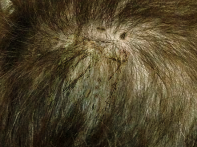
Black Piedra.
Contributed by O Chaigasame, MD
References
Desai DH, Nadkarni NJ. Piedra: an ethnicity-related trichosis? International journal of dermatology. 2014 Aug:53(8):1008-11. doi: 10.1111/j.1365-4632.2012.05722.x. Epub 2013 Jun 20 [PubMed PMID: 23786496]
Level 3 (low-level) evidenceElewski BE, Hazen PG. The superficial mycoses and the dermatophytes. Journal of the American Academy of Dermatology. 1989 Oct:21(4 Pt 1):655-73 [PubMed PMID: 2681280]
Schwartz RA. Superficial fungal infections. Lancet (London, England). 2004 Sep 25-Oct 1:364(9440):1173-82 [PubMed PMID: 15451228]
FISCHMAN O, BLACK PIEDRA IN BRAZIL. A CONTRIBUTION TO ITS STUDY IN MANAUS (STATE OF AMAZONAS). Mycopathologia et mycologia applicata. 1965 Mar 25; [PubMed PMID: 14331326]
Khatu SS, Poojary SA, Nagpur NG. Nodules on the hair: a rare case of mixed piedra. International journal of trichology. 2013 Oct:5(4):220-3. doi: 10.4103/0974-7753.130421. Epub [PubMed PMID: 24778538]
Level 3 (low-level) evidenceKupiec PM, Green AN, Marks KC. Black adherence nodules on the scalp hair shaft. Cutis. 2017 Jul:100(1):14;38;39 [PubMed PMID: 28873102]
Figueras MJ, Guarro J, Zaror L. Ultrastructural aspects of hair digestion in black piedra infection. Journal of medical and veterinary mycology : bi-monthly publication of the International Society for Human and Animal Mycology. 1997 Jan-Feb:35(1):1-6 [PubMed PMID: 9061578]
Level 3 (low-level) evidenceFigueras MJ, Guarro J. X-ray microanalysis of black piedra. Antonie van Leeuwenhoek. 1997 Nov:72(4):275-81 [PubMed PMID: 9442268]
Lacarrubba F, Verzì AE, Micali G. Trichoscopy in the Differential Diagnosis of Pseudonits. Skin appendage disorders. 2019 Apr:5(3):142-145. doi: 10.1159/000493741. Epub 2019 Jan 3 [PubMed PMID: 31049334]
Rudnicka L, Olszewska M, Waśkiel A, Rakowska A. Trichoscopy in Hair Shaft Disorders. Dermatologic clinics. 2018 Oct:36(4):421-430. doi: 10.1016/j.det.2018.05.009. Epub 2018 Aug 16 [PubMed PMID: 30201151]
Rudnicka L, Rakowska A, Kerzeja M, Olszewska M. Hair shafts in trichoscopy: clues for diagnosis of hair and scalp diseases. Dermatologic clinics. 2013 Oct:31(4):695-708, x. doi: 10.1016/j.det.2013.06.007. Epub [PubMed PMID: 24075554]
Gip L. Black piedra: the first case treated with terbinafine (Lamisil). The British journal of dermatology. 1994 Apr:130 Suppl 43():26-8 [PubMed PMID: 8186138]
Level 3 (low-level) evidenceKhandpur S, Reddy BS. Itraconazole therapy for white piedra affecting scalp hair. Journal of the American Academy of Dermatology. 2002 Sep:47(3):415-8 [PubMed PMID: 12196752]
Vijay A, Gupta S, Rawat S, Jain SK. A Rare Case of Coinfection with White Piedra and Pediculosis Capitis. Indian dermatology online journal. 2017 Jul-Aug:8(4):279-280. doi: 10.4103/idoj.IDOJ_353_16. Epub [PubMed PMID: 28761849]
Level 3 (low-level) evidence