Introduction
The frontal bone forms the anterior and superior portion of the skull (see Image. Frontal Skull). At the beginning of life, this bone is separated by a temporary suture called the "frontal suture," which fuses as part of normal development to form the singular frontal bone (see Image. Frontal Bone, Outer Surface). The frontal bone plays a vital role in protecting important neural structures and forms the superior aspect of the orbit.[1] Frontal bone fractures occur in roughly 5% to 15% of all traumatic facial fractures.[2][3]
Clinically, the frontal bone holds significant importance in both medical and surgical contexts. This bone's proximity to the frontal sinus, brain, and orbit means that fractures or infections can result in serious complications, including cerebrospinal fluid leaks, orbital injuries, and intracranial involvement. In aesthetic medicine, the frontal bone also plays a foundational role in forehead contouring and facial rejuvenation procedures. Understanding the anatomy of this bone aids in accurate diagnosis, surgical planning, and prevention of complications.
Structure and Function
Register For Free And Read The Full Article
 Search engine and full access to all medical articles
- 10 free questions in your specialty
- Free CME/CE Activities
- Free daily question in your email
- Save favorite articles to your dashboard
- Emails offering discounts
Learn more about a Subscription to StatPearls Point-of-Care
Structure and Function
The frontal bone forms the anterosuperior part of the cranium and borders several other bones on its external surface. This bone divides into several anatomical regions. The squamous part comprises the superior 2/3 and is commonly referred to as the forehead. A midline prominence called the "glabella" appears in its lower portion.
Just inferior and anterior to the squamous part lies the nasal segment, which joins the nasal bones at the midline to form the nasion. On each side of the nasion, the frontal bone articulates with the frontal processes of the maxilla. The orbital part forms the roof of the orbit and consists of 2 triangular plates extending posteriorly from the supraorbital margins. Lateral to the maxilla, the frontal bone connects with bones forming the posterior orbit, namely, the lacrimal, sphenoid, and zygomatic bones (see Image. Orbit, Anterior View).[4] Posterior and lateral to the zygomatic bones, the frontal bone articulates with the greater wing of the sphenoid and, more posteriorly, with the parietal bones.
On the internal cranial surface, the frontal bone articulates with the ethmoid bone medially and inferiorly and with the sphenoid posteriorly and inferiorly (see Image. Frontal Bone, Inner Surface). Ascending posteriorly and laterally, this bone connects with the temporal bone before meeting the parietal bones at the cranial vertex.
Dense fibrous connective tissue syndesmoses (sutures) between formed bones in the adult calvarium maintain structural integrity.[5] The syndesmosis connecting the frontal bone to the paired parietal bones is called the "coronal suture." Running transversely across the skull’s crown, this suture forms the posterior boundary of the frontal bone. The point where the coronal suture intersects the sagittal suture, uniting the 2 parietal bones, is known as the bregma. In infancy, this region remains open as a nonossified fibrous membrane called the "anterior fontanelle."[6] Laterally, the coronal suture joins the sphenoid, parietal, and temporal bones at the pterion, an anatomically significant H-shaped junction.[7]
The frontal bone contains 3 foramina: a paired set of supraorbital foramina and a singular foramen caecum. The bilateral supraorbital notches lie at the superior orbital rim, just medial to the midpupillary line, transmitting the supraorbital artery, veins, and nerves.[8] The foramen caecum appears on the internal surface near the posterior border, where the frontal bone meets the ethmoid bone, and carries an emissary vein to the superior sagittal sinus. This sinus follows a longitudinal groove running anteroposteriorly along the frontal bone.[9]
The anteroinferior portion of the frontal bone houses 2 frontal sinuses located superior to the orbital roofs. These sinuses vary widely in size and volume between individuals.[10]
Embryology
The frontal bone originates from neural crest cells, along with the anterior cranial bones.[11] At birth, the bone exists as 2 halves separated by the frontal (metopic) suture, which typically fuses during early childhood. In approximately 5% of individuals, the suture remains partially visible beyond infancy, usually just superior to the nasion—a condition known as metopism.[12] The bregma in adults corresponds to the location of the anterior fontanelle in infants. This junction of the coronal, sagittal, and frontal sutures serves as a critical landmark in neonatal assessments. Premature fusion of these sutures may result in craniosynostosis, a developmental abnormality affecting cranial shape (see Image. Frontal Bone at Birth).[13]
Blood Supply and Lymphatics
Blood supply to the head primarily originates from the external and internal carotid arteries (see Image. Arteries of the Scalp and Face). The vertebral arteries contribute vascular supply to the brainstem and form the posterior portion of the circle of Willis. The internal carotid arteries supply the anterior segment of the circle of Willis.[14] The external carotid artery supplies the superficial structures of the head, including the muscles of facial expression and mastication.
Intracranial venous drainage occurs through dural venous sinuses, which converge and ultimately drain into the internal jugular vein. Superficial structures of the head drain primarily via the external jugular vein. Both jugular veins empty into the brachiocephalic vein in the thorax, which then drains into the superior vena cava.
Lymphatic drainage of the head and neck occurs through a network of lymphatic vessels. The right hemicranium drains into the right lymphatic duct, while the left hemicranium drains into the thoracic duct. These ducts then empty into the corresponding right and left subclavian veins.[15]
Nerves
The dermatomal region overlying the frontal bone receives sensory innervation from the ophthalmic branch of the trigeminal nerve (cranial nerve V), which exits the cranium through the paired supraorbital foramina to supply the skin anterior to the frontal bone. Motor innervation to the muscles overlying the frontal bone is provided by the facial nerve (cranial nerve VII). The distribution area includes the frontalis, procerus, and orbicularis oculi muscles, which contribute to facial expression and are clinically relevant in both neurologic and aesthetic evaluations.
Muscles
On the lateral aspect of the frontal bone, just anterior to the parietal bone and superior to the sphenoid bone, the frontal bone contributes to the anterior portion of the temporal fossa. The temporalis muscle occupies this space and is innervated by the trigeminal nerve, playing a key role in jaw closure.[16]
Three muscles associated with the frontal bone are essential for facial expression: the frontalis, procerus, and orbicularis oculi. The frontalis muscles are broad, paired structures that elevate the forehead and eyebrows. The procerus muscles lie superior to the orbit and lateral to the glabella, contributing to frowning. The orbicularis oculi consists of concentric fibers surrounding the eye, enabling blinking and eye closure.[17]
Surgical Considerations
Frontal bone injuries in adults most often result from trauma. A 2014 retrospective study of nearly 4,000 frontal bone fractures identified motorcycle accidents as the leading cause, accounting for approximately 1/3 of cases.[18] Surgical management depends on the location, displacement, and complexity of the fracture, as well as any associated maxillofacial or intracranial injuries. Surgeons may take advantage of preexisting lacerations for access or, when necessary, develop a bicoronal flap to expose the fracture. Fixation typically involves titanium plates with screws, titanium mesh, or a combination of both to achieve stable open reduction and internal fixation.[19]
Potential postoperative complications include surgical site infection, paresthesias of the supraorbital and supratrochlear nerves, cerebrospinal fluid leak, and pneumocephalus. Frontal bone osteomyelitis, although rare, may occur when implanted materials used for fixation become secondarily infected.[20]
Clinical Significance
The anterior fontanelle, which later becomes the bregma, serves as an important landmark during the physical examination of infants. A bulging fontanelle may indicate elevated intracranial pressure and prompt further diagnostic evaluation.[21] Conversely, a sunken fontanelle can signal dehydration. Fontanelles, sutures, and overall cranial shape should be monitored for signs of premature closure or positional abnormalities such as plagiocephaly—a unilateral flattening of the anterior or posterior quadrant of the skull.[22] If left unaddressed, this condition may impair normal brain growth and development.
The pterion, located on the lateral aspect of the frontal bone, is a junction where 4 cranial bones converge: the frontal, temporal, sphenoid, and parietal bones. This region holds clinical importance due to its proximity to the middle meningeal artery, a major vessel supplying the dura mater. Trauma to the pterion warrants urgent evaluation to exclude an epidural hematoma, as arterial injury in this area may result in rapid neurological decline, coma, or death if not managed promptly.[23]
The frontalis muscle is also relevant in aesthetic and reconstructive medicine. Frequent activation of this muscle contributes to horizontal forehead wrinkles, making it a common target for botulinum toxin injection to reduce dynamic lines and improve facial aesthetics.[24]
Frontal bone fractures may arise from facial trauma. Presenting symptoms may include pain, edema, ecchymosis, facial asymmetry, paresthesia, and diplopia.[25]
Media
(Click Image to Enlarge)
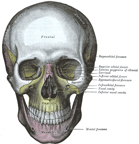
Frontal Skull. This view of the skull allows visualization of the sphenoid, maxilla, frontal, and zygomatic bones.
Henry Vandyke Carter, Public domain, via Wikimedia Commons
(Click Image to Enlarge)
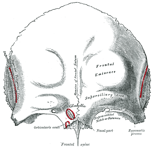
Frontal Bone, Outer Surface. This image shows the anatomical features of the frontal bone, including the glabella, frontal eminence, superciliary arch, frontal spine, remnants of the frontal suture, nasal part, zygomatic process, supraorbital notch, and muscle attachment sites.
Henry Vandyke Carter, Public Domain, via Wikimedia Commons
(Click Image to Enlarge)
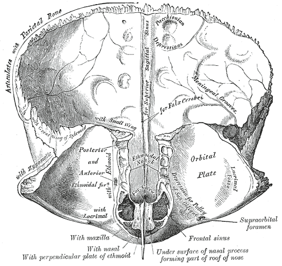
Frontal Bone, Inner Surface. This image shows the inner surface of the frontal bone and its anatomical relationships with adjacent structures.
Henry Vandyke Carter, Public Domain, via Wikimedia Commons
(Click Image to Enlarge)
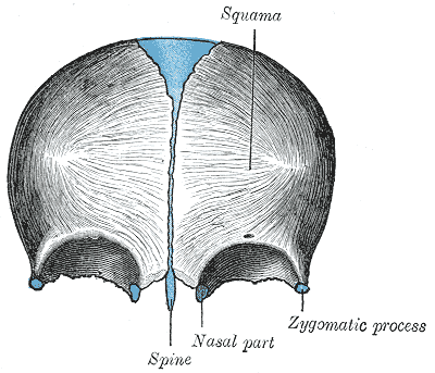
Frontal Bone at Birth. This image shows the squama, spine, nasal part, and zygomatic process of the frontal bone at birth.
Henry Vandyke Carter, Public Domain, via Wikimedia Commons
(Click Image to Enlarge)

Orbit, Anterior View. Shown in this illustration are the supraorbital notch, ethmoidal foramina, optic foramen, superior orbital fissure (hourglass configuration), greater wing of the sphenoid bone, zygomaticofacial foramen, inferior orbital fissure, infraorbital groove, zygomaticomaxillary suture, infraorbital foramen, infraorbital suture, posterior lacrimal crest, anterior lacrimal crest, frontomaxillary suture, and lamina papyracea. The walls of the orbit include the frontal bone superiorly; ethmoid, frontal, lacrimal, and sphenoid bones medially; maxilla, zygomatic, and palatine bones inferiorly; and zygomatic and sphenoid bones laterally.
Johannes Sobotta, MD, Public Domain, Wikimedia Commons
(Click Image to Enlarge)
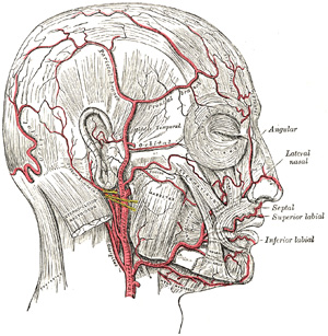
Arteries of the Scalp and Face. Shown here are the branches of the internal and external carotid arteries that supply the scalp and face.
Henry Vandyke Carter, Public Domain, via Wikimedia Commons
References
Singh R. Study of Incidence and Location of Latero-Orbital Foramen Along With Associated Clinical Implications in Adult Indian Dry Skulls. The Journal of craniofacial surgery. 2025 Apr 2:():. doi: 10.1097/SCS.0000000000011350. Epub 2025 Apr 2 [PubMed PMID: 40172975]
Kambalimath DH, Sridhar KR, Achutha S. Surgical Management of Frontal Bone Fractures. The Journal of craniofacial surgery. 2021 Jun 1:32(4):1472-1475. doi: 10.1097/SCS.0000000000007394. Epub [PubMed PMID: 33645950]
Podolsky DJ, Moe KS. Frontal Sinus Fractures. Seminars in plastic surgery. 2021 Nov:35(4):274-283. doi: 10.1055/s-0041-1736325. Epub 2021 Oct 7 [PubMed PMID: 34819810]
Vigo V, Cornejo K, Nunez L, Abla A, Rodriguez Rubio R. Immersive Surgical Anatomy of the Craniometric Points. Cureus. 2020 Jun 15:12(6):e8643. doi: 10.7759/cureus.8643. Epub 2020 Jun 15 [PubMed PMID: 32685312]
Ganz JC. Cranial sutures. Progress in brain research. 2024:285():127-136. doi: 10.1016/bs.pbr.2024.02.019. Epub 2024 Apr 25 [PubMed PMID: 38705712]
D'Antoni AV, Donaldson OI, Schmidt C, Macchi V, De Caro R, Oskouian RJ, Loukas M, Shane Tubbs R. A comprehensive review of the anterior fontanelle: embryology, anatomy, and clinical considerations. Child's nervous system : ChNS : official journal of the International Society for Pediatric Neurosurgery. 2017 Jun:33(6):909-914. doi: 10.1007/s00381-017-3406-1. Epub 2017 Apr 10 [PubMed PMID: 28396968]
Uz A, Korkmaz AC, Filgueira L, Guner MA, Tubbs RS, Demirciler AK. Anatomic Analysis of the Internal and External Aspects of the Pterion. World neurosurgery. 2020 May:137():84-88. doi: 10.1016/j.wneu.2020.01.198. Epub 2020 Feb 3 [PubMed PMID: 32028010]
Voljevica A, Talović E, Šahinović M, Pleho-Kapić A. Morphometric Analysis of the Supraorbital Foramen and Notch in the Population of Bosnia and Herzegovina. Acta medica academica. 2022 Aug:51(2):92-98. doi: 10.5644/ama2006-124.377. Epub [PubMed PMID: 36318001]
Patchana T, Zampella B, Berry JA, Lawandy S, Sweiss RB. Superior Sagittal Sinus: A Review of the History, Surgical Considerations, and Pathology. Cureus. 2019 May 3:11(5):e4597. doi: 10.7759/cureus.4597. Epub 2019 May 3 [PubMed PMID: 31309022]
Dassi CS, Demarco FR, Mangussi-Gomes J, Weber R, Balsalobre L, Stamm AC. The Frontal Sinus and Frontal Recess: Anatomical, Radiological and Surgical Concepts. International archives of otorhinolaryngology. 2020 Jul:24(3):e364-e375. doi: 10.1055/s-0040-1713923. Epub 2020 Jul 31 [PubMed PMID: 32754249]
Anderson BW, Kortz MW, Black AC, Al Kharazi KA. Anatomy, Head and Neck, Skull. StatPearls. 2025 Jan:(): [PubMed PMID: 29763009]
Zdilla MJ, Russell ML, Koons AW, Bliss KN, Mangus KR. Metopism: a Study of the Persistent Metopic Suture. The Journal of craniofacial surgery. 2018 Jan:29(1):204-208. doi: 10.1097/SCS.0000000000004030. Epub [PubMed PMID: 29049140]
Sullivan LE, Li R, Tong VS, Jagasia P, Bonfield CM, Golinko MS, Pontell ME. Craniosynostosis: Current Evaluation and Management. Annals of plastic surgery. 2024 Dec 1:93(6S Suppl 3):S144-S149. doi: 10.1097/SAP.0000000000004131. Epub [PubMed PMID: 39527402]
Menshawi K, Mohr JP, Gutierrez J. A Functional Perspective on the Embryology and Anatomy of the Cerebral Blood Supply. Journal of stroke. 2015 May:17(2):144-58. doi: 10.5853/jos.2015.17.2.144. Epub 2015 May 29 [PubMed PMID: 26060802]
Level 3 (low-level) evidenceLan YL, Wang H, Chen A, Zhang J. Update on the current knowledge of lymphatic drainage system and its emerging roles in glioma management. Immunology. 2023 Feb:168(2):233-247. doi: 10.1111/imm.13517. Epub 2022 Jul 10 [PubMed PMID: 35719015]
Yu SK, Kim TH, Yang KY, Bae CJ, Kim HJ. Morphology of the temporalis muscle focusing on the tendinous attachment onto the coronoid process. Anatomy & cell biology. 2021 Sep 30:54(3):308-314. doi: 10.5115/acb.21.074. Epub [PubMed PMID: 34353976]
Vigliante CE. Anatomy and functions of the muscles of facial expression. Oral and maxillofacial surgery clinics of North America. 2005 Feb:17(1):1-15, v [PubMed PMID: 18088760]
Marinheiro BH, de Medeiros EH, Sverzut CE, Trivellato AE. Frontal bone fractures. The Journal of craniofacial surgery. 2014 Nov:25(6):2139-43. doi: 10.1097/SCS.0000000000001102. Epub [PubMed PMID: 25377971]
Level 2 (mid-level) evidenceSrinivasa R, Furtado SV, Sansgiri T, Vala K. Management of Frontal Bone Fracture in a Tertiary Neurosurgical Care Center-A Retrospective Study. Journal of neurosciences in rural practice. 2022 Jan:13(1):60-66. doi: 10.1055/s-0041-1740615. Epub 2022 Jan 5 [PubMed PMID: 35110921]
Level 2 (mid-level) evidenceJing XL, Luce E. Frontal Sinus Fractures: Management and Complications. Craniomaxillofacial trauma & reconstruction. 2019 Sep:12(3):241-248. doi: 10.1055/s-0038-1675560. Epub 2019 Feb 19 [PubMed PMID: 31428249]
Oumer M, Tazebew A, Alemayehu M. Anterior Fontanel Size Among Term Newborns: A Systematic Review and Meta-Analysis. Public health reviews. 2021:42():1604044. doi: 10.3389/phrs.2021.1604044. Epub 2021 May 10 [PubMed PMID: 34692179]
Level 1 (high-level) evidenceJung BK, Yun IS. Diagnosis and treatment of positional plagiocephaly. Archives of craniofacial surgery. 2020 Apr:21(2):80-86. doi: 10.7181/acfs.2020.00059. Epub 2020 Apr 20 [PubMed PMID: 32380806]
Muche A. Positions and Types of Pterion in Adult Human Skulls: A Preliminary Study. Ethiopian journal of health sciences. 2021 Jul:31(4):875-884. doi: 10.4314/ejhs.v31i4.23. Epub [PubMed PMID: 34703188]
Lee KL, Choi YJ, Gil YC, Hu KS, Tansatit T, Kim HJ. Locational Relationship between the Lateral Border of the Frontalis Muscle and the Superior Temporal Line. Plastic and reconstructive surgery. 2019 Feb:143(2):293e-298e. doi: 10.1097/PRS.0000000000005202. Epub [PubMed PMID: 30489500]
Fakoya AO, Hohman MH, Westbrook KE, Varacallo MA. Anatomy, Head and Neck: Facial Muscles. StatPearls. 2025 Jan:(): [PubMed PMID: 29630261]