Introduction
Striae distensae, commonly known as stretch marks, are a common dermatological concern that can lead to significant cosmetic and psychological distress. They typically develop in areas of the body subject to rapid stretching, including the abdomen, buttocks, thighs, breasts, back, axillae, and groin. These marks are classified according to their appearance or epidemiology as follows:
- Striae atrophicans (characterized by thinned skin)
- Striae gravidarum (following pregnancy)
- Striae rubrae (red in color)
- Striae albae (white in color)
- Striae nigra (black in color)
- Striae caerulea (dark blue in color)
Although striae are benign and asymptomatic, they remain a common concern in both dermatology and aesthetic medicine. For clinicians, it is crucial to differentiate striae from other linear dermatoses (eg, linear morphea or anetoderma) and to be familiar with evidence-based prevention and treatment modalities, ranging from topical therapies and laser treatments to microneedling and emerging regenerative technologies.[1][2]
Etiology
Register For Free And Read The Full Article
 Search engine and full access to all medical articles
- 10 free questions in your specialty
- Free CME/CE Activities
- Free daily question in your email
- Save favorite articles to your dashboard
- Emails offering discounts
Learn more about a Subscription to StatPearls Point-of-Care
Etiology
Striae distensae are a form of dermal scarring resulting from the stretching of the dermis. They often result from rapid weight changes (both gain and loss) or are associated with endogenous or exogenous corticosteroid exposure. Proposed mechanisms include hormonal influences, physical stretching, and structural alterations of dermal collagen and elastic tissue.
Adrenocorticotropic hormones stimulate fibroblast activity and increase protein catabolism. Pregnancy-related hormones may also have a role. Studies have noted lower serum relaxin levels in women with striae distensae.[3] A deficiency of fibrillin has also been suggested as a contributing factor.[4] Although genetic factors remain largely unexplored, decreased expression of collagen and fibronectin genes has been associated with the development of striae.
Epidemiology
Striae distensae are commonly observed in pregnancy (43%–88%), puberty (6%–86%), and obesity (43%). Striae atrophicans are typically associated with underlying medical conditions, particularly Cushing syndrome, and with treatments such as exogenous corticosteroid use (topical or systemic) or surgical interventions.[5] Other associated diseases are Marfan syndrome,[6] anorexia nervosa,[7] various febrile illnesses, and chronic liver disease. Medications associated with the development of striae include chemotherapy agents, prolonged antibiotic use, hormonal contraceptives,[8] and neuroleptics. A positive family history is considered a risk factor for striae.[9]
Striae distensae are more prevalent in females than males and may be more common among certain racial groups. One study found that Black women had a greater number of striae than White women of similar age, parity, weight gain, and family history.[10] Striae may also appear more prominent in individuals with darker skin tones. During pregnancy, younger women are more likely to develop striae compared to older women. Several studies have linked higher prevalence to larger abdominal circumference and significant weight gain, often related to increased fetal size or polyhydramnios.[11]
Another study reported a higher prevalence of striae in smokers compared to nonsmokers. Medical comorbidities such as urinary incontinence and pelvic floor dysfunction have also been associated with the presence of striae.[10][12]
Pathophysiology
The pathophysiology of striae distensae involves disruption of the dermal connective tissue, particularly collagen and elastin fibers, due to mechanical stretching and hormonal influences. Elastases released by mast cells and macrophages are believed to contribute to this process.[13] Rapid skin expansion leads to structural damage in the dermis, while elevated levels of corticosteroids or genetic predisposition can impair fibroblast function and reduce collagen synthesis. Elastolysis in the mid-dermis is followed by reorganization of collagen and fibrillin, leading to dermal atrophy and the formation of characteristic linear, atrophic streaks. Inflammatory mediators are also thought to contribute, particularly in the early stages, accounting for the erythematous appearance of striae rubrae before they evolve into hypopigmented striae albae.
Histopathology
Histopathology of striae rubrae reveals an excess of fine elastic fibers in the papillary dermis, accompanied by thicker, tortuous fibers at the periphery. Perivascular lymphocytic infiltration, dilated dermal vessels, and edema are also present. Elastin and fibrillin fibers show reduction and reorganization, while collagen fibers become thicker and densely packed in parallel rows.
Histopathology of striae albae reveals epidermal atrophy, loss of rete ridges, reduced vascularity, and densely packed, thin, scar-like horizontal collagen bundles.[4] These striae resemble mature atrophic scars. Electron microscopy studies have also demonstrated mast cell degranulation, macrophage activation, and elastolysis in the mid-dermis.[14]
History and Physical
A detailed history often explains the appearance of striae distensae through associations with skin stretching, such as pregnancy, pubertal growth spurts, intense muscular exercise, or weight gain. Inquiring about the recent or past use of potent topical corticosteroids or prolonged systemic steroids is important. If striae are widespread and no obvious cause is identified, obtain a comprehensive medical history and perform a thorough clinical examination.[15]
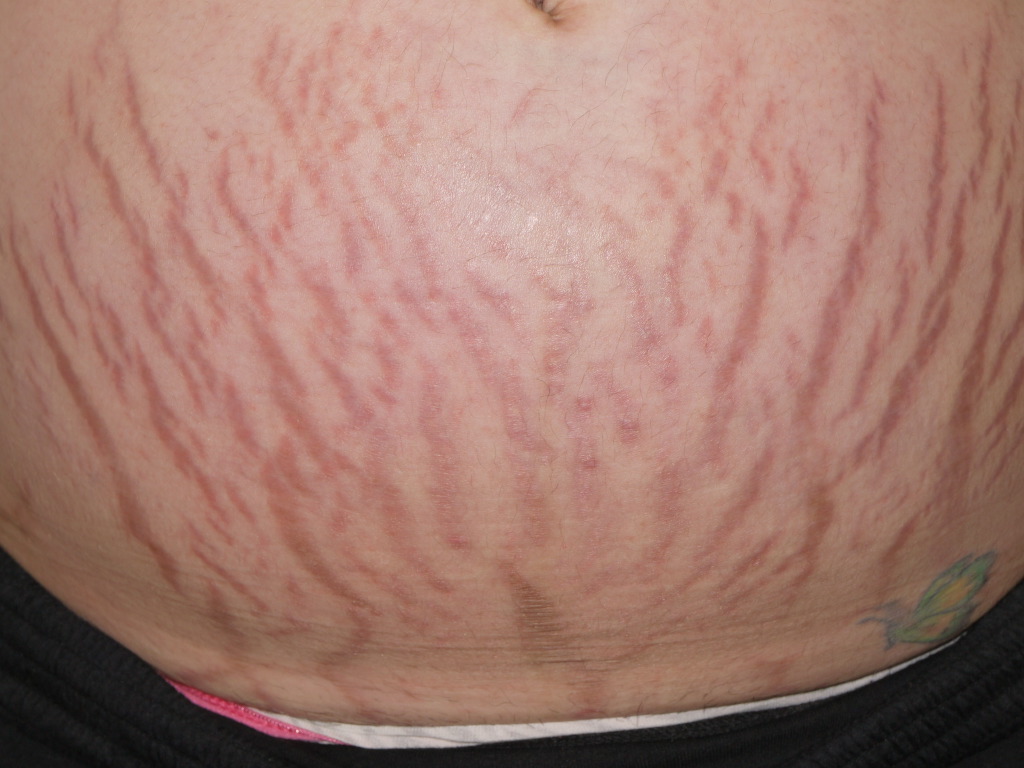
Striae distensae initially present as slightly raised, pink to violaceous linear marks (striae rubrae) that, over months to years, fade into hypopigmented, atrophic, and wrinkled scars (striae albae). These marks typically run perpendicular to the direction of skin tension and gradually fade over time. In pregnancy, striae commonly develop on the abdomen, breasts, and thighs (see Image. Striae Gravidarum Rubrae). In adolescents, they are frequently seen on the thighs, buttocks, and breasts in females, and on the back in males.
Striae rubrae can sometimes be pruritic, but otherwise, striae are typically asymptomatic. Treatment is usually pursued due to their cosmetic appearance.
Evaluation
Various methods have been used to assess the type and severity of striae when evaluating treatment efficacy.[16] However, these approaches are neither standardized nor validated. Dermoscopy reveals increased melanization in striae rubrae and decreased melanization in striae albae.[17] A biopsy is not typically necessary or clinically useful for the evaluation of striae.
Treatment / Management
The primary goals of treatment for striae rubrae are to reduce redness, swelling, and irritation. In striae albae, the focus shifts to stimulating collagen and elastic fiber production, improving skin hydration, and reducing inflammation.[18]
Several comprehensive reviews have noted that topical treatments are frequently recommended for the prevention and management of striae, despite minimal evidence supporting their effectiveness. Most clinical trials conducted to date have been of low quality and involved small sample sizes.
-
Despite limited evidence, many emollients and over-the-counter cosmeceuticals are marketed to and commonly used by pregnant women to prevent or reduce the severity of striae distensae. This often involves significant effort and expense, despite the lack of clear evidence supporting their effectiveness.[19]
-
Silicone gels, commonly recommended for atrophic scars, are sometimes used to treat striae distensae. However, published results on their effectiveness remain difficult to interpret.[20]
- Tretinoin cream has shown potential benefit in improving striae rubrae compared to placebo over a 6-month period. However, it may cause irritation, redness, and peeling, and is contraindicated during pregnancy due to its associated risk category.[21][22] (A1)
- Chemical peels with various acids have been used to treat striae, but their effectiveness remains uncertain.
Although physical treatments are sometimes recommended to treat striae, little evidence supports their effectiveness.
- Exposure to broadband ultraviolet radiation can induce repigmentation in striae albae; however, this effect is typically temporary, with loss of pigmentation within a few months.
- Light and laser therapies have demonstrated improvements in the appearance of striae; however, the optimal type of therapy and the most effective stage for treatment (rubrae or albae) remain uncertain.[23] Several laser types, such as pulsed dye lasers, target vascular chromophores in striae rubrae and have been reported to reduce redness and swelling.[24] Fractional lasers, including Erbium-YAG, stimulate fibroblast activity to increase collagen and elastin production and may also promote repigmentation in striae albae.[25][26] (A1)
- Light and laser treatments can cause short-term erythema and edema. In individuals with darker skin types, these therapies carry a higher risk of adverse effects, including transient postinflammatory hyperpigmentation and persistent hypopigmentation. Nonablative devices are generally considered safer than ablative lasers for these patients.
- Radiofrequency energy devices deliver high-frequency alternating current to generate dermal heat, which leads to skin tightening, wrinkle reduction, and improved appearance of cellulite. These treatment approaches promote neocollagenesis, neoelastogenesis, and an increase in ground substances (eg, proteoglycans), thereby theoretically improving the appearance of striae. At least one published study reported that the treatment was well-tolerated and that patients were satisfied with the results. A recent advancement involves delivering radiofrequency energy to a depth of 3.5 mm using a multiple-needle delivery system. While anecdotal reports are encouraging, definitive studies are still lacking.[27][28] (A1)
- Other devices used for treatment include microdermabrasion,[29] galvanopuncture,[30] needling,[31] pulsed magnetic fields,[27] and ultrasound devices.[32][33] (A1)
Assessing the effectiveness of treatments is challenging due to variations in laser protocols, which differ in devices, fluence, pulse duration, spot size, treatment frequency, and number of sessions. The benefits of combination therapies also remain unclear.[18]
Platelet-rich plasma (PRP) injections are also under investigation as a potential treatment that promotes collagen production and tissue regeneration by releasing growth factors.[34] Although some studies report modest improvements in skin texture and appearance, high-quality evidence is limited, and PRP should be considered an adjunct rather than a first-line therapy. When using any of these modalities, obtaining standardized pretreatment and posttreatment photographs is important. Additionally, photographs should be taken at 6 and 12 months after the treatment course. Many published reports lack standardized imaging and long-term follow-up, limiting the assessment of treatment efficacy. (B2)
Differential Diagnosis
When evaluating striae distensae, it is important to consider a range of differential diagnoses that can present with similar linear or atrophic skin changes. Accurate identification is crucial for appropriate management and to exclude underlying systemic or dermatological conditions.
Differential diagnoses include anetoderma, cutis laxa, linear focal elastosis, mid-dermal elastosis, and pseudoxanthoma elasticum.
Prognosis
Striae distensae generally have a favorable prognosis, as they are benign and pose no health risk. Over time, striae typically fade from striae rubrae to striae albae, becoming less noticeable, although they rarely disappear completely. Although their appearance may improve over time or with certain cosmetic treatments, effective long-term resolution is uncommon. Psychosocial distress can persist, especially when striae are extensive or located in visible areas. Thus, prognosis often depends as much on the cosmetic and emotional impact as on the physical presence of the striae.
Complications
Striae distensae are primarily a cosmetic concern and rarely cause physical complications. However, they can have significant psychological and emotional effects, including reduced self-esteem, body image dissatisfaction, and social anxiety. In rare cases, aggressive or inappropriate treatments—such as overuse of topical steroids, invasive procedures, or unregulated cosmetic products—may result in skin irritation, scarring, or infection. Additionally, striae can occasionally signal underlying endocrine disorders, such as Cushing syndrome, warranting further evaluation. Therefore, careful assessment and cautious treatment planning are essential to prevent harm and address any underlying conditions.
Consultations
Management of striae may involve consultation with dermatologists for expert evaluation, diagnosis, and discussion of advanced treatment options such as laser therapy or microneedling. Endocrinologists may be consulted when striae are atypical or extensive or suggest an underlying hormonal disorder, such as Cushing syndrome. Mental health professionals can provide valuable support for patients experiencing significant psychological distress related to striae. An interprofessional approach ensures comprehensive care that addresses both the physical and emotional needs of the patient.
Deterrence and Patient Education
Deterrence and patient education for striae distensae should focus on addressing modifiable risk factors and promoting evidence-based preventive strategies. Physicians should counsel patients on the importance of gradual weight changes, adequate hydration, and a nutrient-rich diet, particularly vitamins C and E, to support skin health and dermal elasticity. Caution is warranted when prescribing topical or systemic corticosteroids, with clear instructions on their appropriate use to avoid misuse that can lead to dermal atrophy. Although no intervention has been definitively proven to prevent striae, recommending regular moisturization and sun protection may help maintain skin integrity. Clear, evidence-based communication is essential to manage patient expectations and discourage the use of ineffective or potentially harmful treatments.
Enhancing Healthcare Team Outcomes
Effective management of striae distensae requires a coordinated, patient-centered approach involving physicians, advanced practitioners, nurses, pharmacists, and other healthcare professionals. Clinicians must have strong diagnostic skills to differentiate striae from other dermatoses and identify underlying causes, such as endocrine disorders, that may require further evaluation. Patient education should highlight the benign nature of striae, the limited effectiveness of current treatments, and the potential risks linked to cosmetic interventions. Ethical care involves transparent communication, avoiding unnecessary or overly aggressive treatments, and promoting informed, shared decision-making.
Nurses play a crucial role in reinforcing patient education and monitoring concerns, whereas pharmacists provide guidance on the appropriate use of topical agents and caution against unproven or harmful products. Effective interprofessional communication among healthcare providers is crucial to deliver consistent messaging, avoid redundant or conflicting advice, and coordinate care when specialty referrals, such as dermatology, endocrinology, or behavioral health, are necessary. By collaborating across disciplines, the healthcare team can enhance patient safety, minimize unnecessary interventions, address psychosocial effects, and improve both individual outcomes and overall team performance.
Media
(Click Image to Enlarge)
References
Farahnik B, Park K, Kroumpouzos G, Murase J. Striae gravidarum: Risk factors, prevention, and management. International journal of women's dermatology. 2017 Jun:3(2):77-85. doi: 10.1016/j.ijwd.2016.11.001. Epub 2016 Dec 6 [PubMed PMID: 28560300]
Al-Himdani S, Ud-Din S, Gilmore S, Bayat A. Striae distensae: a comprehensive review and evidence-based evaluation of prophylaxis and treatment. The British journal of dermatology. 2014 Mar:170(3):527-47. doi: 10.1111/bjd.12681. Epub [PubMed PMID: 24125059]
Level 1 (high-level) evidenceLurie S, Matas Z, Fux A, Golan A, Sadan O. Association of serum relaxin with striae gravidarum in pregnant women. Archives of gynecology and obstetrics. 2011 Feb:283(2):219-22. doi: 10.1007/s00404-009-1332-5. Epub 2010 Jan 3 [PubMed PMID: 20047054]
Wang F, Calderone K, Smith NR, Do TT, Helfrich YR, Johnson TR, Kang S, Voorhees JJ, Fisher GJ. Marked disruption and aberrant regulation of elastic fibres in early striae gravidarum. The British journal of dermatology. 2015 Dec:173(6):1420-30. doi: 10.1111/bjd.14027. Epub 2015 Nov 8 [PubMed PMID: 26179468]
Neve S, Kirtschig G. Elastotic striae associated with striae distensae after application of very potent topical corticosteroids. Clinical and experimental dermatology. 2006 May:31(3):461-2 [PubMed PMID: 16681607]
Level 3 (low-level) evidenceLedoux M, Beauchet A, Fermanian C, Boileau C, Jondeau G, Saiag P. A case-control study of cutaneous signs in adult patients with Marfan disease: diagnostic value of striae. Journal of the American Academy of Dermatology. 2011 Feb:64(2):290-5. doi: 10.1016/j.jaad.2010.01.032. Epub 2010 Nov 26 [PubMed PMID: 21112669]
Level 2 (mid-level) evidenceStrumia R. Skin signs in anorexia nervosa. Dermato-endocrinology. 2009 Sep:1(5):268-70 [PubMed PMID: 20808514]
Gupta M. Medroxyprogesterone acetate [Depo Provera] injections. Development of striae. The British journal of family planning. 2000 Apr:26(2):104-5 [PubMed PMID: 10773604]
Level 3 (low-level) evidenceTürkmen H, Yörük S. Risk factors of striae gravidarum and chloasma melasma and their effects on quality of life. Journal of cosmetic dermatology. 2023 Feb:22(2):603-612. doi: 10.1111/jocd.14783. Epub 2022 Feb 7 [PubMed PMID: 35037372]
Level 2 (mid-level) evidenceElbuluk N, Saizan AL, Hurtado ACM, Hamilton T, Kang S. Differences in clinical features and risk factors for striae distensae in Black and White women. Archives of dermatological research. 2025 Mar 18:317(1):592. doi: 10.1007/s00403-025-04050-z. Epub 2025 Mar 18 [PubMed PMID: 40100381]
Picard D, Sellier S, Houivet E, Marpeau L, Fournet P, Thobois B, Bénichou J, Joly P. Incidence and risk factors for striae gravidarum. Journal of the American Academy of Dermatology. 2015 Oct:73(4):699-700. doi: 10.1016/j.jaad.2015.06.037. Epub [PubMed PMID: 26369842]
Yousefi F, Abbaspoor Z, Siahkal SF, Mohaghegh Z, Ghanbari S, Zahedian M. Association Between Striae and Pelvic Organ Prolapse in Women: A Systematic Review and Meta-Analysis. International urogynecology journal. 2024 Aug:35(8):1561-1570. doi: 10.1007/s00192-024-05832-1. Epub 2024 Jun 12 [PubMed PMID: 38864859]
Level 1 (high-level) evidenceSheu HM, Yu HS, Chang CH. Mast cell degranulation and elastolysis in the early stage of striae distensae. Journal of cutaneous pathology. 1991 Dec:18(6):410-6 [PubMed PMID: 1774350]
Zheng P, Lavker RM, Kligman AM. Anatomy of striae. The British journal of dermatology. 1985 Feb:112(2):185-93 [PubMed PMID: 3970840]
Kasielska-Trojan A, Sobczak M, Antoszewski B. Risk factors of striae gravidarum. International journal of cosmetic science. 2015 Apr:37(2):236-40. doi: 10.1111/ics.12188. Epub 2015 Jan 12 [PubMed PMID: 25440082]
La Padula S, Hersant B, Pizza C, Chesné C, Jamin A, Ben Mosbah I, D'Andrea F, Persichetti P, Rega U, Pensato R, Meningaud JP. The Objective Stretch Marks Photonumeric Assessment Scale: A New and Complete Method to Assess Striae Distensae: Correction. Plastic and reconstructive surgery. 2025 May 1:155(5):905. doi: 10.1097/PRS.0000000000012091. Epub 2025 Apr 23 [PubMed PMID: 40294321]
Hermanns JF, Piérard GE. High-resolution epiluminescence colorimetry of striae distensae. Journal of the European Academy of Dermatology and Venereology : JEADV. 2006 Mar:20(3):282-7 [PubMed PMID: 16503888]
Forbat E, Al-Niaimi F. Treatment of striae distensae: An evidence-based approach. Journal of cosmetic and laser therapy : official publication of the European Society for Laser Dermatology. 2019:21(1):49-57. doi: 10.1080/14764172.2017.1418515. Epub 2018 Feb 16 [PubMed PMID: 29451986]
Rawlings AV, Bielfeldt S, Lombard KJ. A review of the effects of moisturizers on the appearance of scars and striae. International journal of cosmetic science. 2012 Dec:34(6):519-24. doi: 10.1111/j.1468-2494.2012.00751.x. Epub 2012 Sep 21 [PubMed PMID: 22994859]
Ud-Din S, McAnelly SL, Bowring A, Whiteside S, Morris J, Chaudhry I, Bayat A. A double-blind controlled clinical trial assessing the effect of topical gels on striae distensae (stretch marks): a non-invasive imaging, morphological and immunohistochemical study. Archives of dermatological research. 2013 Sep:305(7):603-17. doi: 10.1007/s00403-013-1336-7. Epub 2013 Apr 12 [PubMed PMID: 23579949]
Level 1 (high-level) evidenceUd-Din S, McGeorge D, Bayat A. Topical management of striae distensae (stretch marks): prevention and therapy of striae rubrae and albae. Journal of the European Academy of Dermatology and Venereology : JEADV. 2016 Feb:30(2):211-22. doi: 10.1111/jdv.13223. Epub 2015 Oct 20 [PubMed PMID: 26486318]
Korgavkar K, Wang F. Stretch marks during pregnancy: a review of topical prevention. The British journal of dermatology. 2015 Mar:172(3):606-15. doi: 10.1111/bjd.13426. Epub 2015 Feb 8 [PubMed PMID: 25255817]
Timur Taşhan S, Kafkasli A. The effect of bitter almond oil and massaging on striae gravidarum in primiparaous women. Journal of clinical nursing. 2012 Jun:21(11-12):1570-6. doi: 10.1111/j.1365-2702.2012.04087.x. Epub [PubMed PMID: 22594386]
Level 2 (mid-level) evidenceElsaie ML, Hussein MS, Tawfik AA, Emam HM, Badawi MA, Fawzy MM, Shokeir HA. Comparison of the effectiveness of two fluences using long-pulsed Nd:YAG laser in the treatment of striae distensae. Histological and morphometric evaluation. Lasers in medical science. 2016 Dec:31(9):1845-1853 [PubMed PMID: 27595152]
Zaleski-Larsen LA, Jones IT, Guiha I, Wu DC, Goldman MP. A Comparison Study of the Nonablative Fractional 1565-nm Er: glass and the Picosecond Fractional 1064/532-nm Nd: YAG Lasers in the Treatment of Striae Alba: A Split Body Double-Blinded Trial. Dermatologic surgery : official publication for American Society for Dermatologic Surgery [et al.]. 2018 Oct:44(10):1311-1316. doi: 10.1097/DSS.0000000000001555. Epub [PubMed PMID: 29746426]
Level 1 (high-level) evidenceLozano SH, Gulmatico-Flores Z, Abad-Casintahan MF. A Comparative Study of Picosecond Fractional 1064-nm Nd:YAG Laser Versus Fractional 10,600-nm Carbon Dioxide Laser in the Treatment of Abdominal Striae Alba: A Randomized, Prospective, Assessor-blinded, Split-abdomen Trial. Journal of cosmetic and laser therapy : official publication of the European Society for Laser Dermatology. 2025 Apr 24:():1-8. doi: 10.1080/14764172.2025.2497416. Epub 2025 Apr 24 [PubMed PMID: 40272374]
Level 1 (high-level) evidenceDover JS, Rothaus K, Gold MH. Evaluation of safety and patient subjective efficacy of using radiofrequency and pulsed magnetic fields for the treatment of striae (stretch marks). The Journal of clinical and aesthetic dermatology. 2014 Sep:7(9):30-3 [PubMed PMID: 25276274]
Aktoz F, Yilmaz N. Comparing fractional microneedle radiofrequency and fractional CO2 laser for striae distensae treatment: a systematic review and meta-analysis. Lasers in medical science. 2024 Nov 9:39(1):271. doi: 10.1007/s10103-024-04231-8. Epub 2024 Nov 9 [PubMed PMID: 39516426]
Level 1 (high-level) evidenceHersant B, Niddam J, Meningaud JP. Comparison between the efficacy and safety of platelet-rich plasma vs microdermabrasion in the treatment of striae distensae: clinical and histopathological study. Journal of cosmetic dermatology. 2016 Dec:15(4):565. doi: 10.1111/jocd.12246. Epub 2016 Jun 20 [PubMed PMID: 27320781]
Bitencourt S, Lunardelli A, Amaral RH, Dias HB, Boschi ES, de Oliveira JR. Safety and patient subjective efficacy of using galvanopuncture for the treatment of striae distensae. Journal of cosmetic dermatology. 2016 Dec:15(4):393-398. doi: 10.1111/jocd.12222. Epub 2016 Apr 19 [PubMed PMID: 27090205]
Aust M, Walezko N. [Acne scars and striae distensae: Effective treatment with medical skin needling]. Der Hautarzt; Zeitschrift fur Dermatologie, Venerologie, und verwandte Gebiete. 2015 Oct:66(10):748-52. doi: 10.1007/s00105-015-3662-5. Epub [PubMed PMID: 26251169]
Kravvas G, Veitch D, Al-Niaimi F. The use of energy devices in the treatment of striae: a systematic literature review. The Journal of dermatological treatment. 2019 May:30(3):294-302. doi: 10.1080/09546634.2018.1506078. Epub 2018 Sep 7 [PubMed PMID: 30049244]
Level 1 (high-level) evidenceMustafa A, Zahid R, Khan S, Faisal M, Hassan Farooq MA, Imran J, Malik W, Khan A, Azeem B, Maryam A, Kumar S, Khatri M. Evaluating CO2 laser and micro-needling therapies for striae distensae: a comprehensive meta-analysis and systematic review. Lasers in medical science. 2025 Mar 25:40(1):161. doi: 10.1007/s10103-025-04420-z. Epub 2025 Mar 25 [PubMed PMID: 40131559]
Level 1 (high-level) evidenceGamil HD, Ibrahim SA, Ebrahim HM, Albalat W. Platelet-Rich Plasma Versus Tretinoin in Treatment of Striae Distensae: A Comparative Study. Dermatologic surgery : official publication for American Society for Dermatologic Surgery [et al.]. 2018 May:44(5):697-704. doi: 10.1097/DSS.0000000000001408. Epub [PubMed PMID: 29701622]
Level 2 (mid-level) evidence