Introduction
Perioral dermatitis is a benign eruption that occurs most commonly in young, female adults, consisting of small inflammatory papules and pustules or pink, scaly patches around the mouth. Although the perioral region is the most common distribution area, this disease can also affect the periocular and paranasal skin (see Image. Perioral Dermatitis). For this reason, this dermatologic phenomenon is often referred to as periorificial dermatitis.
A strong association exists between perioral dermatitis and topical corticosteroid use, particularly on the face. Initially responsive to steroids, the condition often worsens with withdrawal, potentially leading to chronic, recurrent disease or a granulomatous variant. Other potential triggers include inhaled or nasal corticosteroids, fluorinated toothpaste, chewing gum, dental materials, cosmetics, sunscreens, facemask use, and improper CPAP therapy. Hormonal factors may also play a role.
Diagnosis of perioral dermatitis is typically clinical, but biopsy or additional testing may be warranted in atypical cases. Despite existing guidelines, clinicians often misdiagnose perioral dermatitis and inappropriately prescribe topical corticosteroids, which worsen the condition over time. Therefore, recognizing steroid-induced flares is essential to reduce iatrogenic harm and ensure effective management.
Topical steroid use on the face can trigger perioral dermatitis, and therefore, a primary recommendation for treatment is the discontinuation of steroid application by the patient. However, Abrupt cessation of corticosteroids can cause rebound flaring; therefore, a gradual taper may be necessary. Other treatment approaches include topical metronidazole, topical calcineurin inhibitors, and oral tetracycline antibiotics. While treatment is generally effective, perioral dermatitis can be persistent or recurrent, requiring long-term management strategies tailored to the individual.
Etiology
Register For Free And Read The Full Article
 Search engine and full access to all medical articles
- 10 free questions in your specialty
- Free CME/CE Activities
- Free daily question in your email
- Save favorite articles to your dashboard
- Emails offering discounts
Learn more about a Subscription to StatPearls Point-of-Care
Etiology
The exact cause of perioral dermatitis is unknown; however, multiple environmental exposures have been suggested as possible underlying etiologies of this condition.
Corticosteroids and Perioral Dermatitis
For many patients, an association between topical corticosteroid use and perioral dermatitis is often noted.[1] Topical steroid use may precede the eruption, and chronic use of topical steroids increases the risk of developing severe disease.[2] Initially, the facial eruption is responsive to the topical steroid use. However, upon withdrawal of the topical steroid, the eruption recurs. This leads to long-term dependency on topical steroid use.[3] Eventually, perioral dermatitis may progress in severity with chronic steroid use and may evolve into a granulomatous subtype of the disease.
In addition to topical steroid use, perioral dermatitis has been reported to occur with the use of nasal and inhaled corticosteroids.[4][5] The exact mechanism through which topical steroids predispose patients to perioral dermatitis is not understood. Authors have postulated that topical steroids influence the microflora of the hair follicle, which may contribute to the pathogenesis of this condition.[6][7][8] Some investigators have proposed infectious sources as a cause for perioral dermatitis, including Candida albicans, fusiform bacteria, and Demodex mites.[6][7][8]
Additional Potential Etiologies
Fluorinated toothpaste use has also been associated with perioral dermatitis.[9] Additionally, chewing gum and dental fillings have been associated with perioral dermatitis.[10][11] Certain cosmetic products, eg, the combined use of moisturizers and foundations, as well as physical sunscreens, have been identified as underlying etiologies in some patients.[12][13]
Given the female predominance with this condition, hormonal factors have been considered in the etiology. Interestingly, oral contraceptive pills have been associated with the improvement of perioral dermatitis.[14] In recent years, anti-COVID-19 facemasks have also been linked to the development of perioral dermatitis.[15] Additionally, improper use of activated oxygen in CPAP has also been linked to the development of perioral dermatitis.[16]
Epidemiology
Perioral dermatitis commonly affects young adult females, 20 to 45 years of age.[17][18] However, it has also been reported in children.[19] No significant difference in the frequency of perioral dermatitis among gender or race in pediatric populations has been observed.[20]
Pathophysiology
The clinical manifestations of perioral dermatitis occur when perifollicular and perivascular inflammation lead to erythematous papules around the mouth, eyes, and nose. However, the exact pathophysiological mechanism is unknown. The underlying etiology is likely multifactorial with contributions from genetic, hormonal, and environmental factors.[21]
Histopathology
A skin biopsy of perioral dermatitis reveals a perifollicular and perivascular lymphohistiocytic inflammatory infiltrate with sparse plasma cells. Although follicular spongiosis may be present, typical dermatitis features are often absent despite this term being used to name the disease.[22] A granulomatous variant of perioral dermatitis has been identified in which dermal epithelioid granulomas and giant cells are observed, in addition to the perivascular and perifollicular lymphohistiocytic inflammation.[23][24]
History and Physical
Perioral dermatitis is primarily clinically diagnosed. Patients with perioral dermatitis most commonly present with characteristic erythematous grouped papules that are often bilateral but may be unilateral, surrounding the mouth, eyes, and nose. Since it frequently affects the skin surrounding other facial orifices, this condition has also been termed periorificial dermatitis. Other clinical findings associated with perioral dermatitis include scaling, vesicles, and pustules. The vermilion borders of the lips are typically spared of involvement.
In the granulomatous variant, flesh-colored to erythematous to yellow-brown papules erupt in the same distribution. However, rare involvement of the ears, neck, scalp, trunk, vulva, and extremities has been described.[25][26] Patients often report that the areas of involvement are associated with symptoms of burning or sensitivity, although pruritus can also occur.[27] Patients may also mention sensitivity to various skincare products, which may exacerbate the rash.[28]
Additionally, rare cases of granulomatous periorificial dermatitis may have concomitant blepharitis or conjunctivitis; therefore, an ophthalmologic evaluation should also be considered. Other systemic manifestations are not associated with this disease.[26] Lupoid perioral dermatitis is a more severe variant of perioral dermatitis that presents with dense groupings of red-brown papules. Lupoid perioral dermatitis lesions demonstrate a lupoid infiltrate on diascopy.[29]
Evaluation
The vast majority of patients with perioral dermatitis are diagnosed clinically, although skin biopsy should be considered with an atypical presentation or lack of response to treatment. If a bacterial component is suspected, a culture may be obtained. Additionally, a scraping and potassium hydroxide (KOH) prep can be done if Candida is considered to be contributing to the lesions. With a more severe disease presentation, diascopy can be performed by an experienced clinician.
Treatment / Management
Topical Therapies
First-line treatment options for perioral dermatitis include metronidazole cream or gel, clindamycin lotion or gel, erythromycin gel, topical sulfur preparations, and azelaic acid gel.[30][31][32] In the treatment of this condition, antibiotics are helpful for their anti-inflammatory properties.[33] Topical calcineurin inhibitors, eg, tacrolimus ointment or pimecrolimus cream, can also be effective.[34][35][36][37] Topical sulfur or sulfacetamide preparations, and topical adapalene have been demonstrated to show improvement.[38][39] Photodynamic therapy using 5-aminolevulinic acid as a photosensitizer has been reported to provide benefit.[40](A1)
Oral Treatment Approaches
If topical therapies are not helpful, or for extensive involvement of perioral dermatitis, oral antibiotics are often helpful. Tetracycline 250 to 500 mg twice a day, doxycycline 100 mg twice a day or once a day, and minocycline 100 mg twice a day or once a day, for an 8 to 12 week tapering course can be prescribed. When tetracycline antibiotics are contraindicated, as they are in children younger than 8, nursing mothers, and pregnant females, erythromycin 250 mg to 500 mg daily can be used as an alternative treatment.[41][42] (B3)
The goal of oral antibiotic therapy is to provide rapid improvement, but topical therapies should be used concurrently. Topical therapies may not show peak efficacy until 3 months of daily treatment. Therefore, the objective is to stop oral antibiotics if possible after 3 months. However, a subset of patients may require longer courses of oral antibiotics if they cannot be controlled with chronic daily topical therapies. In recalcitrant and severe cases, low-dose oral isotretinoin can be used, initially at 0.2 mg/kg per day, then tapering to 0.1 to 0.05 mg/kg per day.[43][44](B3)
Corticosteroid Considerations
Since perioral dermatitis is not a primary eczematous process, recognizing that topical corticosteroids should not be used is important. Although topical corticosteroids may provide a temporary benefit, the rash can often flare and worsen when they are discontinued.
If patients have been using topical corticosteroids to treat the eruption, abrupt discontinuation may lead to rebound flaring, and patients should be forewarned that the condition will likely worsen until it improves with the initiation of appropriate therapies for perioral dermatitis. If a patient has been using a medium to high potency steroid, it may need to be slowly weaned by using a low potency steroid (eg, hydrocortisone cream).[45] Treatment of granulomatous perioral dermatitis with 1.5% topical ruxolitinib cream has recently shown promising results.[46](B3)
Differential Diagnosis
The differential diagnoses of perioral dermatitis include:
- Rosacea: Rosacea presents as inflammatory papules and pustules, primarily of the central face, including the nose. Patients often have accompanying telangiectatic erythema with flushing. Many authors view perioral dermatitis as a variant of rosacea, as the 2 conditions typically respond to the same therapies. Clinically, this is distinguished from acne vulgaris by the lack of comedones, which is characteristic of acne.[33]
- Acne vulgaris: This presents as inflammatory papules, pustules, cysts, and comedones primarily on the face, but may also affect the chest and back. Patients may have acne flares, eg, a helmet chin strap, secondary to mechanical mechanisms. The distribution of involvement may mimic perioral dermatitis. Adult female acne is characterized by inflammatory papules of the chin and jawline and can have a similar distribution to perioral dermatitis.
- Sarcoidosis: Sarcoidosis can also manifest features similar to rosacea and perioral dermatitis with red-brown papules on the periorificial regions of the face. Typically, sarcoidosis lesions are more widespread and can occur in other skin locations, and patients often have symptoms of systemic sarcoidosis. A biopsy is helpful in distinguishing sarcoidosis.
- Seborrheic dermatitis: Seborrheic dermatitis presents as ill-defined erythematous patches with greasy scale distributed on the eyebrows, glabella, paranasal skin, nasolabial folds, beard, scalp, and chest.
- Allergic contact dermatitis: This condition can also be considered but tends to present with ill-defined scaling macules, patches, and plaques with possible lichenification. If patients with perioral dermatitis do not improve with conventional treatments, patch testing should be considered to rule out allergic contact dermatitis from skincare or oral care products.
- Irritant cheilitis: Irritant cheilitis, eg, lip licker's cheilitis, presents with erythema and scaling of the cutaneous lip. This typically also affects the vermilion lip and does not spare the vermilion border. In contrast, the vermilion border is spared in perioral dermatitis.
- Demodex folliculitis: This condition presents with scattered erythematous facial papules and pustules. Demodex mites have been implicated in the pathogenesis of rosacea. Unroofing of the pustules, followed by microscopy of the purulent material, shows numerous Demodex mites.
- Tinea faciei: This dermatologic condition presents with erythematous scaling papules and annular plaques and can be ruled out by performing a KOH prep and microscopic examination of the scale.
- Cutaneous adnexal neoplasms: Syringomas and other cutaneous adnexal neoplasms can also mimic perioral dermatitis, as these lesions are flesh-colored to erythematous facial papules. A biopsy is helpful for diagnosis.
Prognosis
In some patients, the rash may resolve completely after discontinuation of an offending agent, including topical steroids and skincare products. Frequently, however, perioral dermatitis can be a chronic relapsing condition and often requires long-term treatment.
Complications
Complications of perioral dermatitis include:
- Emotional distress due to the chronicity of the disease
- Poor quality of life due to sometimes disfiguring lesions on the face
- Scarring may occur with the lupoid variant [29]
Consultations
If symptoms of conjunctivitis, eg, burning, itching, or tearing, are present, an ophthalmology consultation may be warranted.
Deterrence and Patient Education
Deterrence and patient education play a vital role in effectively managing perioral dermatitis. Patients should be clearly informed about potential triggers, especially the harmful impact of topical corticosteroids on facial skin, including over-the-counter products. Clinicians must emphasize discontinuing all topical steroids and avoiding other potential irritants such as fluorinated toothpaste, heavy cosmetics, and unnecessary skincare products.
Patients should be instructed to use only the prescribed treatment regimen and understand that a temporary worsening of symptoms—known as a rebound flare—may occur after stopping steroids. Education should also include realistic expectations regarding the treatment timeline, which may span several weeks to months, and the possibility of chronic or recurrent symptoms. Open communication and consistent reinforcement of these points can significantly improve adherence and long-term outcomes.
Enhancing Healthcare Team Outcomes
Effective management of perioral dermatitis requires a collaborative, patient-centered approach involving physicians, advanced practitioners, nurses, pharmacists, and other healthcare professionals. Primary care providers and dermatologists are typically responsible for diagnosing and initiating treatment, but optimal outcomes depend on a coordinated effort across the care team. Physicians and advanced practitioners must evaluate the severity of disease, choose appropriate therapies, and provide patients with realistic expectations, especially regarding the potential for rebound flares after discontinuing topical corticosteroids. Nurses and medical assistants play a crucial role in patient education, emphasizing adherence to prescribed regimens, monitoring for adverse effects, and ensuring patients understand the importance of follow-up care. Pharmacists contribute by reviewing prescriptions for potential drug interactions, counseling patients on proper medication use, and advising on adverse effects, particularly those associated with tetracycline antibiotics, eg, photosensitivity, gastrointestinal upset, and allergic reactions.
Strong interprofessional communication is essential to enhance patient safety, care coordination, and overall team performance. Open channels between all members of the healthcare team help ensure that any concerns—such as intolerance to medications or unexpected adverse effects—are addressed promptly. Nurses and pharmacists should be empowered to alert prescribers to issues that may necessitate a change in therapy or additional patient follow-up. Additionally, patients should be encouraged to report any new or worsening symptoms to the care team without delay. Regular case reviews, shared documentation, and a unified care plan help avoid miscommunication and reinforce consistent messaging to the patient. This collaborative approach not only improves treatment adherence and outcomes but also builds trust and fosters a more responsive, patient-centered care environment.
Media
(Click Image to Enlarge)
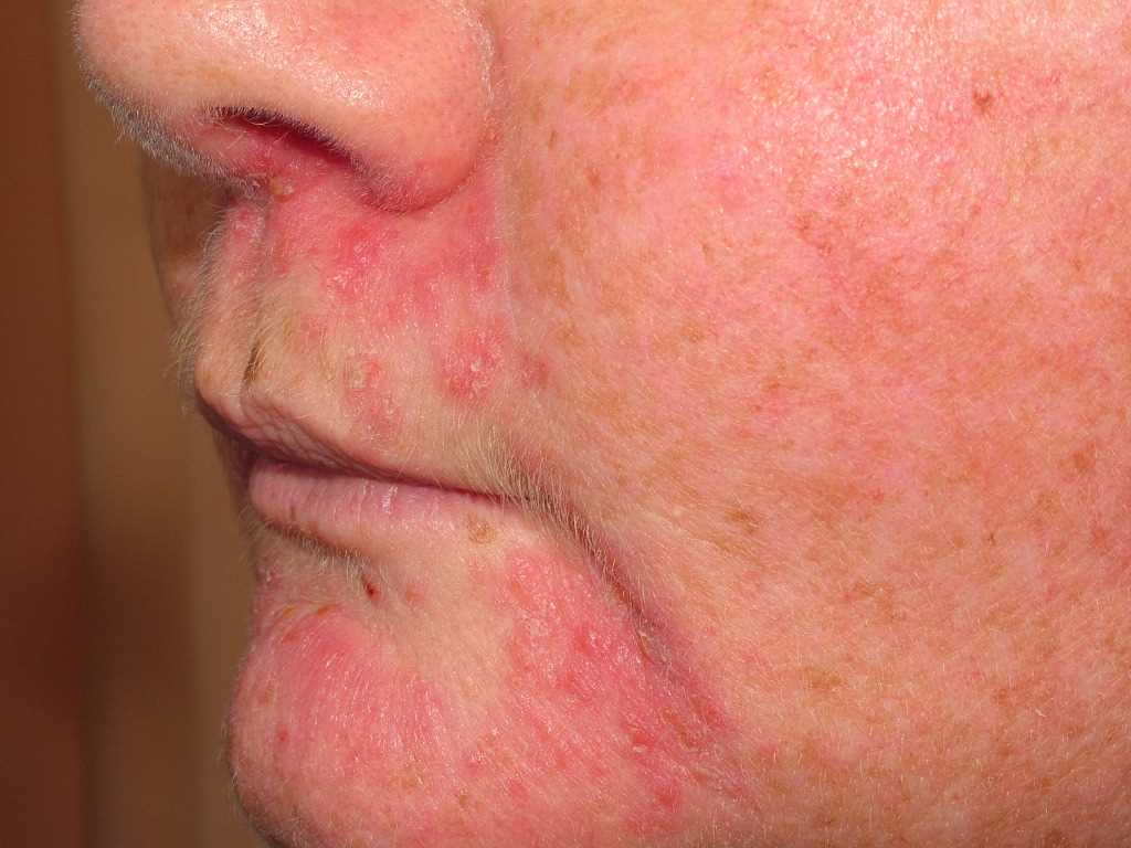
Perioral Dermatitis, Perioral dermatitis is a benign eruption that occurs most commonly in young, female adults, consisting of small inflammatory papules and pustules or pink, scaly patches around the mouth.
References
Kosari P, Feldman SR. Case report: Fluocinonide-induced perioral dermatitis in a patient with psoriasis. Dermatology online journal. 2009 Mar 15:15(3):15 [PubMed PMID: 19379659]
Level 3 (low-level) evidenceLjubojeviae S, Basta-Juzbasiae A, Lipozenèiae J. Steroid dermatitis resembling rosacea: aetiopathogenesis and treatment. Journal of the European Academy of Dermatology and Venereology : JEADV. 2002 Mar:16(2):121-6 [PubMed PMID: 12046812]
Hengge UR, Ruzicka T, Schwartz RA, Cork MJ. Adverse effects of topical glucocorticosteroids. Journal of the American Academy of Dermatology. 2006 Jan:54(1):1-15; quiz 16-8 [PubMed PMID: 16384751]
Peralta L, Morais P. Perioral dermatitis -- the role of nasal steroids. Cutaneous and ocular toxicology. 2012 Jun:31(2):160-3. doi: 10.3109/15569527.2011.621918. Epub 2011 Oct 13 [PubMed PMID: 21995785]
Level 3 (low-level) evidencePoulos GA, Brodell RT. Perioral dermatitis associated with an inhaled corticosteroid. Archives of dermatology. 2007 Nov:143(11):1460 [PubMed PMID: 18025385]
Level 3 (low-level) evidenceBradford LG, Montes LF. Perioral dermatitis and Candida albicans. Archives of dermatology. 1972 Jun:105(6):892-5 [PubMed PMID: 4555327]
Takiwaki H, Tsuda H, Arase S, Takeichi H. Differences between intrafollicular microorganism profiles in perioral and seborrhoeic dermatitis. Clinical and experimental dermatology. 2003 Sep:28(5):531-4 [PubMed PMID: 12950346]
Dolenc-Voljc M, Pohar M, Lunder T. Density of Demodex folliculorum in perioral dermatitis. Acta dermato-venereologica. 2005:85(3):211-5 [PubMed PMID: 16040404]
Level 3 (low-level) evidencePeters P, Drummond C. Perioral dermatitis from high fluoride dentifrice: a case report and review of literature. Australian dental journal. 2013 Sep:58(3):371-2. doi: 10.1111/adj.12077. Epub [PubMed PMID: 23981221]
Level 3 (low-level) evidenceSatyawan I, Oranje AP, van Joost T. Perioral dermatitis in a child due to rosin in chewing gum. Contact dermatitis. 1990 Mar:22(3):182-3 [PubMed PMID: 2335094]
Level 3 (low-level) evidenceGuarneri F, Marini H. Perioral dermatitis after dental filling in a 12-year-old girl: involvement of cholinergic system in skin neuroinflammation? TheScientificWorldJournal. 2008 Feb 6:8():157-63. doi: 10.1100/tsw.2008.31. Epub 2008 Feb 6 [PubMed PMID: 18264633]
Level 3 (low-level) evidenceMalik R, Quirk CJ. Topical applications and perioral dermatitis. The Australasian journal of dermatology. 2000 Feb:41(1):34-8 [PubMed PMID: 10715898]
Level 2 (mid-level) evidenceAbeck D, Geisenfelder B, Brandt O. Physical sunscreens with high sun protection factor may cause perioral dermatitis in children. Journal der Deutschen Dermatologischen Gesellschaft = Journal of the German Society of Dermatology : JDDG. 2009 Aug:7(8):701-3. doi: 10.1111/j.1610-0387.2009.07045.x. Epub 2009 Feb 23 [PubMed PMID: 19250246]
Spirov G, Berova N, Vassilev D. Effect of oral inhibitors of ovulation in treatment of rosacea and dermatitis perioralis in women. The Australasian journal of dermatology. 1971 Dec:12():145-54 [PubMed PMID: 12305771]
Level 3 (low-level) evidenceVeraldi S, Mattioli MA, Nazzaro G. Anti-COVID-19 Face Masks and Perioral Dermatitis. The Journal of clinical and aesthetic dermatology. 2023 May:16(5):22 [PubMed PMID: 37288278]
Ahmed G, Pal S, Esquinas AM, Karim HMR. Perioral Dermatitis Caused by Improper Use of Activated Oxygen in CPAP users and its Complexity. The Journal of clinical and aesthetic dermatology. 2022 Aug:15(8):14 [PubMed PMID: 36061482]
Hogan DJ, Epstein JD, Lane PR. Perioral dermatitis: an uncommon condition? CMAJ : Canadian Medical Association journal = journal de l'Association medicale canadienne. 1986 May 1:134(9):1025-8 [PubMed PMID: 2938708]
Lipozencic J, Ljubojevic S. Perioral dermatitis. Clinics in dermatology. 2011 Mar-Apr:29(2):157-61. doi: 10.1016/j.clindermatol.2010.09.007. Epub [PubMed PMID: 21396555]
Manders SM, Lucky AW. Perioral dermatitis in childhood. Journal of the American Academy of Dermatology. 1992 Nov:27(5 Pt 1):688-92 [PubMed PMID: 1430388]
Level 3 (low-level) evidenceLaude TA, Salvemini JN. Perioral dermatitis in children. Seminars in cutaneous medicine and surgery. 1999 Sep:18(3):206-9 [PubMed PMID: 10468040]
Tempark T, Shwayder TA. Perioral dermatitis: a review of the condition with special attention to treatment options. American journal of clinical dermatology. 2014 Apr:15(2):101-13. doi: 10.1007/s40257-014-0067-7. Epub [PubMed PMID: 24623018]
Marks R, Black MM. Perioral dermatitis. A histopathologic study of 26 cases. The British journal of dermatology. 1971 Mar:84(3):242-7 [PubMed PMID: 5572676]
Level 3 (low-level) evidenceFrieden IJ, Prose NS, Fletcher V, Turner ML. Granulomatous perioral dermatitis in children. Archives of dermatology. 1989 Mar:125(3):369-73 [PubMed PMID: 2923443]
Level 3 (low-level) evidenceRamelet AA, Delacrétaz J. [Histopathologic study of perioral dermatitis]. Dermatologica. 1981:163(5):361-9 [PubMed PMID: 7333393]
Nguyen V, Eichenfield LF. Periorificial dermatitis in children and adolescents. Journal of the American Academy of Dermatology. 2006 Nov:55(5):781-5 [PubMed PMID: 17052482]
Level 2 (mid-level) evidenceUrbatsch AJ, Frieden I, Williams ML, Elewski BE, Mancini AJ, Paller AS. Extrafacial and generalized granulomatous periorificial dermatitis. Archives of dermatology. 2002 Oct:138(10):1354-8 [PubMed PMID: 12374542]
Level 3 (low-level) evidenceKuflik JH, Janniger CK, Piela Z. Perioral dermatitis: an acneiform eruption. Cutis. 2001 Jan:67(1):21-2 [PubMed PMID: 11204598]
Wilkinson DS, Kirton V, Wilkinson JD. Perioral dermatitis: a 12-year review. The British journal of dermatology. 1979 Sep:101(3):245-57 [PubMed PMID: 159711]
Baratli J, Megahed M. [Lupoid perioral dermatitis as a special form of perioral dermatitis: review of pathogenesis and new therapeutic options]. Der Hautarzt; Zeitschrift fur Dermatologie, Venerologie, und verwandte Gebiete. 2013 Dec:64(12):888-90. doi: 10.1007/s00105-013-2684-0. Epub [PubMed PMID: 24201654]
Level 3 (low-level) evidenceVeien NK, Munkvad JM, Nielsen AO, Niordson AM, Stahl D, Thormann J. Topical metronidazole in the treatment of perioral dermatitis. Journal of the American Academy of Dermatology. 1991 Feb:24(2 Pt 1):258-60 [PubMed PMID: 2007672]
Level 1 (high-level) evidenceMiller SR, Shalita AR. Topical metronidazole gel (0.75%) for the treatment of perioral dermatitis in children. Journal of the American Academy of Dermatology. 1994 Nov:31(5 Pt 2):847-8 [PubMed PMID: 7962733]
Level 3 (low-level) evidenceJansen T. Azelaic acid as a new treatment for perioral dermatitis: results from an open study. The British journal of dermatology. 2004 Oct:151(4):933-4 [PubMed PMID: 15491447]
Level 3 (low-level) evidenceHoepfner A, Marsela E, Clanner-Engelshofen BM, Horvath ON, Sardy M, French LE, Reinholz M. Rosacea and perioral dermatitis: a single-center retrospective analysis of the clinical presentation of 1032 patients. Journal der Deutschen Dermatologischen Gesellschaft = Journal of the German Society of Dermatology : JDDG. 2020 Jun:18(6):561-570. doi: 10.1111/ddg.14120. Epub 2020 May 29 [PubMed PMID: 32469453]
Level 2 (mid-level) evidenceGoldman D. Tacrolimus ointment for the treatment of steroid-induced rosacea: a preliminary report. Journal of the American Academy of Dermatology. 2001 Jun:44(6):995-8 [PubMed PMID: 11369912]
Level 3 (low-level) evidenceChu CY. The use of 1% pimecrolimus cream for the treatment of steroid-induced rosacea. The British journal of dermatology. 2005 Feb:152(2):396-9 [PubMed PMID: 15727676]
Level 3 (low-level) evidenceGerber PA, Neumann NJ, Ruzicka T, Bruch-Gerharz D. [Perioral dermatitis following treatment with tacrolimus]. Der Hautarzt; Zeitschrift fur Dermatologie, Venerologie, und verwandte Gebiete. 2005 Oct:56(10):967-8 [PubMed PMID: 16142496]
Level 3 (low-level) evidenceSchwarz T, Kreiselmaier I, Bieber T, Thaci D, Simon JC, Meurer M, Werfel T, Zuberbier T, Luger TA, Wollenberg A, Bräutigam M. A randomized, double-blind, vehicle-controlled study of 1% pimecrolimus cream in adult patients with perioral dermatitis. Journal of the American Academy of Dermatology. 2008 Jul:59(1):34-40. doi: 10.1016/j.jaad.2008.03.043. Epub 2008 May 7 [PubMed PMID: 18462835]
Level 1 (high-level) evidenceBendl BJ. Perioral dermatitis: etiology and treatment. Cutis. 1976 May:17(5):903-8 [PubMed PMID: 1035149]
Level 1 (high-level) evidenceJansen T. Perioral dermatitis successfully treated with topical adapalene. Journal of the European Academy of Dermatology and Venereology : JEADV. 2002 Mar:16(2):175-7 [PubMed PMID: 12046830]
Level 3 (low-level) evidenceRichey DF, Hopson B. Photodynamic therapy for perioral dermatitis. Journal of drugs in dermatology : JDD. 2006 Feb:5(2 Suppl):12-6 [PubMed PMID: 16485876]
Choi YL, Lee KJ, Cho HJ, Kim WS, Lee JH, Yang JM, Lee ES, Lee DY. Case of childhood granulomatous periorificial dermatitis in a Korean boy treated by oral erythromycin. The Journal of dermatology. 2006 Nov:33(11):806-8 [PubMed PMID: 17073999]
Level 3 (low-level) evidenceEllis CN, Stawiski MA. The treatment of perioral dermatitis, acne rosacea, and seborrheic dermatitis. The Medical clinics of North America. 1982 Jul:66(4):819-30 [PubMed PMID: 6212726]
Smith KW. Perioral dermatitis with histopathologic features of granulomatous rosacea: successful treatment with isotretinoin. Cutis. 1990 Nov:46(5):413-5 [PubMed PMID: 2148143]
Level 3 (low-level) evidenceLipozenčić J, Hadžavdić SL. Perioral dermatitis. Clinics in dermatology. 2014 Jan-Feb:32(1):125-30. doi: 10.1016/j.clindermatol.2013.05.034. Epub [PubMed PMID: 24314386]
Searle T, Ali FR, Al-Niaimi F. Perioral dermatitis: Diagnosis, proposed etiologies, and management. Journal of cosmetic dermatology. 2021 Dec:20(12):3839-3848. doi: 10.1111/jocd.14060. Epub 2021 Mar 16 [PubMed PMID: 33751778]
Tran SS, Ungar B, Brunner PM. Treatment of granulomatous perioral dermatitis with 1.5% topical ruxolitinib cream. JAAD case reports. 2024 May:47():1-3. doi: 10.1016/j.jdcr.2024.02.022. Epub 2024 Mar 6 [PubMed PMID: 38576899]
Level 3 (low-level) evidence