Introduction
Flail chest is a traumatic injury of the thorax that involves fractures of 3 or more ribs in at least 2 places (see Image. Three-Dimensional Reconstruction of Flail Chest). Although this definition is widely used, clinical findings show that even smaller segments involving 1 or 2 ribs can function as flail segments if they move independently and generate negative intrapleural pressure. The Western Trauma Association, in its rib fracture algorithm, describes this anatomical pattern as a disconnected portion of the chest wall that moves paradoxically inward during inspiration. Flail chest is fundamentally a clinical diagnosis, as not all patients with this fracture pattern exhibit clinical signs of paradoxical motion. This injury disrupts normal respiratory mechanics and imposes a greater physiological burden on older patients and those with chronic pulmonary disease.
Flail chest frequently results from severe blunt thoracic trauma and often occurs along with other high-impact injuries. Pain and associated trauma both contribute to the complexity of treatment. Although most cases are unilateral, bilateral involvement can occur. Radiographic findings may suggest flail chest, but clinical observation of paradoxical movement confirms the diagnosis.
Etiology
Register For Free And Read The Full Article
 Search engine and full access to all medical articles
- 10 free questions in your specialty
- Free CME/CE Activities
- Free daily question in your email
- Save favorite articles to your dashboard
- Emails offering discounts
Learn more about a Subscription to StatPearls Point-of-Care
Etiology
As a traumatic disorder, flail chest shares risk factors with other forms of major trauma. Male sex and intoxication are independent risk factors. Motor vehicle collisions account for approximately 75% of major trauma cases leading to flail chest, while falls, particularly among older adults, cause an additional 15%.[1] Certain high-impact injuries, such as direct blows to the thorax, are more likely to produce 2 fractures on a single rib, increasing the likelihood of a flail segment. In contrast, rollover and crush injuries tend to cause single-point rib fractures and are, therefore, less likely to result in flail chest.
Metabolic bone disorders, such as osteogenesis imperfecta, increase susceptibility in pediatric populations. Among older adults, age-related stiffening of the chest wall and underlying osteoporosis contribute to increased risk. Chronic pulmonary conditions, which are prevalent in this group, also heighten vulnerability to complications associated with flail chest.
Epidemiology
According to the American Association for the Surgery of Trauma, approximately 1% of the United States population sustains a significant traumatic injury each year. Chest trauma occurs in 20% of these cases and accounts for 25% of trauma-related deaths. Flail chest is identified in roughly 7% of patients with chest trauma.[2] Hospitalization is typically required, and flail chest occurs in isolation in fewer than 40% of cases. More commonly, this traumatic condition is accompanied by pulmonary contusions, hemothorax or pneumothorax, head injury, and occasionally major vascular injury. Reported mortality ranges from 10% to 20%, often attributable to associated injuries rather than flail chest itself.[3] Morbidity is substantial due to prolonged hospitalization and complex recovery.
Pathophysiology
Airflow into and out of the lungs depends on changes in intrathoracic pressure. Inspiration requires coordinated activation of respiratory muscle groups, including the diaphragm, external intercostals, parasternal muscles, internal intercostals, and accessory muscles. Descent of the diaphragmatic dome increases the vertical dimension of the thoracic cavity and generates negative pressure. At rest, the diaphragm alone can sustain adequate ventilation. During exercise or in pathological states, the intercostal muscles play a significant role in generating inspiratory effort. Exhalation is typically passive due to the elastic recoil of the lungs, although the abdominal and intercostal muscles may assist.
In flail chest, disruption of chest wall continuity impairs the normal biomechanical function of the ribs. The flail segment moves in paradoxical opposition to the remainder of the thoracic cage, collapsing inward during inspiration as the intact chest wall expands outward. The severity of this paradoxical motion and its physiological impact is determined by 3 factors: pleural pressure, the extent of the flail segment, and intercostal muscle activation during inspiration.[4]
A flail segment disrupts normal respiratory mechanics in 3 principal ways: ineffective ventilation, pulmonary contusion, and hypoventilation with atelectasis. Ventilation becomes ineffective due to increased dead space, reduced intrathoracic pressure, and elevated oxygen demand from injured tissues. Pulmonary contusion in the lung adjacent to the flail segment is nearly universal and results in edema, hemorrhage, and, in some cases, tissue necrosis. These changes impair gas exchange and reduce pulmonary compliance.
Hypoventilation and atelectasis arise from pain associated with the injury. Pain induces both voluntary and involuntary splinting, in which patients limit chest wall movement to avoid discomfort. This restriction leads to shallow respiration, reduced tidal volume, and an ineffective cough, all of which contribute to atelectasis, impaired gas exchange in the affected lung region, and retention of pulmonary secretions.[5]
History and Physical
The history is often evident, as flail chest typically occurs in the context of major blunt force trauma. Older adults are at increased risk. Diagnosis may be more challenging in nonverbal patients, victims of abuse, or cases without a reliable history.
The physical examination should be performed according to a systematic trauma assessment in all patients with suspected thoracic injury. The patient should be fully exposed. A complete set of vital signs should be obtained, including an accurate respiratory rate and oxygen saturation. The primary survey, comprising airway, breathing, circulation, disability, and exposure (eg, ABCDE), should be initiated, followed by the secondary survey. Bilateral breath sounds should be auscultated, and the chest should be palpated for tenderness, deformity, or crepitus. The thorax should be inspected for bruising, open wounds, or seatbelt marks. Patients typically report severe chest wall pain and may present with tachypnea, splinting, or signs of respiratory insufficiency.
Flail chest is characterized by localized pain, pronounced tenderness over the fracture site, and respiratory distress, including tachypnea and dyspnea. Paradoxical chest wall motion, when visible, is a key diagnostic sign. During inspiration, the flail segment moves inward as the rest of the chest expands. During expiration, the segment moves outward as the chest contracts. However, this pathognomonic finding may be absent, especially in patients with significant splinting and shallow respirations. Paradoxical motion may become more evident as intercostal muscle fatigue progresses. Positive pressure ventilation alters thoracic pressure dynamics and eliminates paradoxical movement. Consequently, patients on bilevel positive airway pressure or mechanical ventilation do not exhibit this sign, and the diagnosis of flail chest may only become apparent following extubation.
Evaluation
Evaluation of a patient with trauma should include all standard examinations and studies used in the assessment of individuals with major multisystem trauma. The extended focused assessment with sonography in trauma may detect pneumothorax or hemothorax, but does not typically aid in the identification of flail chest. Chest radiography is often the first imaging study that raises suspicion for flail chest. However, even with 2 views, the sensitivity of this modality is limited, and rib fractures may be missed. A rib series, which includes posteroanterior and oblique views, is more sensitive than standard chest radiographs but may still fail to detect up to one-fourth of rib fractures.[6] Relying solely on chest radiography may result in missed diagnoses.
Computed tomography (CT) is the imaging test of choice for evaluating flail chest and associated injuries. CT with 3-dimensional reconstruction provides more detailed visualization of fracture patterns and aids in clinical correlation. The finding of 3 ribs fractured in 2 locations is suggestive of a flail segment, but must be assessed in the context of clinical signs. CT imaging may also guide therapeutic decisions. Trauma laboratory testing should include arterial blood gas analysis. While laboratory studies do not directly diagnose flail chest, they are essential for monitoring the progression of evolving respiratory failure.
Treatment / Management
Nonoperative management of flail chest is guided by 5 key principles: pain control, respiratory support, pulmonary hygiene, fluid balance, and the avoidance of outdated interventions. These principles aim to restore respiratory function, reduce the need for surgical intervention, and minimize complications. Effective analgesia, particularly with regional techniques such as thoracic epidural or paravertebral blocks, is essential not only for patient comfort but also for preventing respiratory compromise. Multimodal regimens decrease opioid requirements and improve pulmonary outcomes.
Respiratory support encompasses supplemental oxygen, noninvasive ventilation (NIV), and, when necessary, invasive mechanical ventilation, using lung-protective strategies. NIV may prevent the need for intubation in selected patients, but it must be closely monitored due to the risk of failure. Pulmonary hygiene relies on deep breathing exercises, physiotherapy, and early mobilization to optimize secretion clearance and reduce the risk of atelectasis and pneumonia. Preserving mucociliary function and preventing secretion retention are essential for avoiding pulmonary complications.
Fluid management requires judicious resuscitation. Isotonic fluids may be used initially, but excessive hydration can worsen pulmonary contusion and contribute to acute respiratory distress syndrome. Obsolete interventions, such as external taping or corticosteroid administration, are no longer recommended. Prehospital care emphasizes stabilization of airway, breathing, and circulation while avoiding high-pressure ventilation that could aggravate pneumothorax. Early, coordinated interprofessional care is crucial for maintaining physiological stability, preventing complications, and minimizing the need for surgical intervention.[7](B2)
Mechanical ventilation using positive pressure, originally termed “internal pneumatic stabilization” by Avery et al in 1956, is a primary intervention for patients with flail chest complicated by respiratory failure or concomitant head or abdominal trauma requiring ventilatory support. This approach restores synchronous movement between the flail segment and the intact chest wall, thereby improving respiratory mechanics. Advancements, such as volume-limited ventilation, have contributed to a decrease in mortality in severe cases. However, prolonged mechanical ventilation poses a significant risk for ventilator-associated pneumonia, highlighting the importance of careful patient selection, close monitoring, and timely weaning.[8][9](A1)
Surgical stabilization of rib fractures (SSRF) is currently recognized as a key treatment option for patients with severe thoracic injury, particularly those with flail chest. Possible approaches include open reduction and internal fixation, minimally invasive methods such as video-assisted thoracoscopic surgery, and percutaneous procedures. These modalities aim to restore chest wall stability, alleviate pain, and improve respiratory function.
Indications and Contraindications for Surgical Stabilization of Rib Fractures
The decision to perform SSRF is guided by an assessment of the patient's clinical condition, fracture pattern, associated injuries, and facility capabilities. The 2024 position paper by the World Society of Emergency Surgery and Chest Wall Injury Society identifies key indications for SSRF. These indications include flail chest with respiratory compromise or prolonged ventilator dependence, particularly when extubation fails after 7 to 14 days, and significant chest wall deformity or volume loss exceeding 30% on imaging.
Other indications include intractable pain unresponsive to optimal nonoperative measures, symptomatic nonunion or malunion resulting in chronic pain or disability, and the opportunity to stabilize fractures during thoracotomy performed for other intrathoracic procedures. SSRF is also recommended in cases of multiple (≥3) ipsilateral displaced rib fractures associated with respiratory dysfunction, even in the absence of a flail segment, as well as in settings where pulmonary herniation is due to chest wall defects.[10]
Absolute contraindications include hemodynamic instability, rib fractures outside ribs 3 to 10 (with limited exceptions), acute myocardial infarction, and, traditionally, severe traumatic brain injury (TBI). However, emerging evidence supports the use of SSRF in select cases of TBI. Relative contraindications include being younger than 18, having substantial comorbidities, mild to moderate TBI or spinal cord injury, pleural empyema, prior chest wall irradiation, extensive soft tissue loss, and inability to position the patient safely for surgery.
Timing of Surgical Stabilization of Rib Fractures
Early surgery (within 48–72 hours) is associated with better outcomes, including a shorter duration of mechanical ventilation, a limited intensive care unit and hospital length of stay, a lower incidence of pneumonia, and a decreased need for tracheostomy. Delayed surgery (>72 hours) is less advantageous and technically more difficult, though it may still be appropriate for addressing chronic complications such as nonunion. Operating during the peak of inflammation (>72 hours) may lead to poorer outcomes.[11][12][13][14][15](A1)
Surgical Technique and Hardware
Surgical planning is guided by detailed imaging, ideally using 3-dimensional CT reconstructions. SSRF is performed under general anesthesia, often with single-lung ventilation. Incisions are designed to spare muscle tissue, and intraoperative ultrasound may assist in accurately localizing target fractures. Not all fractures require fixation. Sufficient stability is often achieved by addressing only the most displaced ribs.
Fixation equipment options include the following:
- Bicortical screw plating systems, typically precontoured titanium plates with locking screw options (eg, MatrixRib, RibFix Blu)
- U-plate systems (eg, RibLoc U Plus), which offer stronger constructs and wider load distribution
- Intramedullary devices (eg, splints, K-wires) for fractures not amenable to plating
- Bioresorbable implants, now less frequently used due to high early failure rates
- Judet struts, largely obsolete today [16]
Outcomes improve when fixation is limited to fractures contributing most to instability. This approach facilitates faster recovery and fewer complications.
Postoperative Care
Surgical wounds are closed in layers, and a chest tube is placed when the pleural cavity is entered. Perioperative analgesia may be provided using thoracic epidural catheters.
Differential Diagnosis
The following conditions should be considered in the differential diagnosis of flail chest:
- Acute aortic dissection
- Amebiasis
- Pediatric trauma considerations, such as pulmonary contusion without rib fracture, nonaccidental trauma, and congenital chest wall anomalies
- Domestic violence
- Suspected abuse in older adults
- Esophagitis
- Clavicle fracture
- Mechanical back pain
- Pneumothorax
- Pulmonary embolism
- Sternal fracture
- Upper genitourinary trauma
These conditions reflect a broad spectrum of thoracic and systemic conditions that can complicate the clinical picture in suspected flail chest cases. Accurate differentiation prevents delays in diagnosis and optimizes patient outcomes.
Prognosis
Patients who do not require mechanical ventilation generally have a better prognosis than those who do. However, adverse events are common and often result in prolonged disability, with pain persisting for months or even years in some cases.
Complications
Flail chest can lead to the following complications:
- Severe chronic pain
- Chest wall deformity
- Dyspnea
- Loss of exercise endurance
The complications of flail chest extend beyond the acute phase and can cause lasting morbidity. Early recognition and comprehensive care are essential to optimize recovery.
Deterrence and Patient Education
Preventing traumatic injuries such as flail chest begins with implementing safety measures such as wearing seatbelts and using protective gear during high-risk activities. Public education campaigns can raise awareness about fall prevention and safe driving. Prompt assessment and early treatment of chest trauma should be emphasized to all healthcare professionals involved in emergency and trauma care, including paramedics, emergency clinicians, surgeons, and nurses. This information should also be communicated to the general public to raise awareness about the importance of seeking immediate medical attention for chest trauma.
Pearls and Other Issues
As mentioned, flail chest is associated with significant complications. Consequently, this condition requires a high index of suspicion. The diagnosis is primarily clinical and most often occurs when at least 3 ribs are fractured in at least 2 places, though not all such injuries result in flail chest.
Older adults are particularly vulnerable to flail chest and its complications. Children rarely develop flail chest due to the elasticity of their chest wall. However, the occurrence of flail chest in this population indicates severe trauma and warrants thorough evaluation. Mechanical ventilation is reserved for cases of respiratory failure and should be avoided whenever possible. NIV is preferred when clinically appropriate to prevent intubation.
Enhancing Healthcare Team Outcomes
Flail chest constitutes a significant traumatic injury with the potential for considerable morbidity. Optimal management requires a well-coordinated, interprofessional team approach, supported by clear communication among all involved disciplines. Essential components of effective care, such as providing adequate ventilatory support, achieving optimal analgesia, and maintaining pulmonary hygiene, demand collaborative efforts from various healthcare professionals to secure favorable clinical outcomes.[17][18]
Most patients require admission, and some may need intensive care. Nurses caring for patients with flail chest must be aware of likely complications and know when to consult the managing clinician. Respiratory therapy is typically involved in assisting with coughing, clearing secretions, and maintaining airway patency. Pain specialists are essential, as most patients experience moderate to severe pain.
Dietitians should be involved to ensure adequate nutrition and prevent muscle wasting. Oxygenation may be compromised, with some patients requiring mechanical ventilation. Others may warrant surgery to stabilize fractured rib segments. These patients often have prolonged hospital stays, and nurses must ensure the appropriate administration of deep vein thrombosis and stress ulcer prophylaxis. Lastly, physical therapy is crucial in preventing muscle loss and contractures. Only through such an interprofessional team approach can morbidity from this condition be minimized.
Outcomes
Data indicate that flail chest, accompanied by other organ injuries, carries a mortality rate of 5% to 10%. Complications may include acute respiratory distress syndrome, sepsis, respiratory failure, and pneumonia. Overall, patients who do not require mechanical ventilation experience better outcomes than those who are intubated. Even without additional organ injury, recovery from flail chest is often prolonged. Most patients require 6 to 12 months to recover fully, and some continue to experience chest pain for many years.[19]
Media
(Click Image to Enlarge)
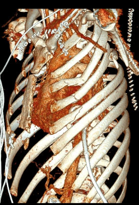
Three-Dimensional Reconstruction of Flail Chest. Multiple rib fractures create a flail segment with abnormal chest wall motion. The 3-dimensional image highlights the extent of injury.
Contributed by Yvonne M. Carter, MD
References
Bastos R, Calhoon JH, Baisden CE. Flail chest and pulmonary contusion. Seminars in thoracic and cardiovascular surgery. 2008 Spring:20(1):39-45. doi: 10.1053/j.semtcvs.2008.01.004. Epub [PubMed PMID: 18420125]
Dehghan N, de Mestral C, McKee MD, Schemitsch EH, Nathens A. Flail chest injuries: a review of outcomes and treatment practices from the National Trauma Data Bank. The journal of trauma and acute care surgery. 2014 Feb:76(2):462-8. doi: 10.1097/TA.0000000000000086. Epub [PubMed PMID: 24458051]
Level 2 (mid-level) evidenceBenjamin E, Recinos G, Aiolfi A, Inaba K, Demetriades D. Flail Chest: Less Deadly than Originally Thought. World journal of surgery. 2018 Dec:42(12):3927-3931. doi: 10.1007/s00268-018-4723-6. Epub [PubMed PMID: 29922874]
Poirier WJ, Vacca VM Jr. Flail chest. Nursing. 2013 Dec:43(12):10-1. doi: 10.1097/01.NURSE.0000437477.45498.8a. Epub [PubMed PMID: 24257520]
Level 3 (low-level) evidenceRogers FB, Larson NJ, Rhone A, Amaya D, Olson-Bullis BA, Blondeau BX. Comprehensive Review of Current Pain Management in Rib Fractures With Practical Guidelines for Clinicians. Journal of intensive care medicine. 2023 Apr:38(4):327-339. doi: 10.1177/08850666221148644. Epub 2023 Jan 4 [PubMed PMID: 36600614]
Chardoli M, Hasan-Ghaliaee T, Akbari H, Rahimi-Movaghar V. Accuracy of chest radiography versus chest computed tomography in hemodynamically stable patients with blunt chest trauma. Chinese journal of traumatology = Zhonghua chuang shang za zhi. 2013:16(6):351-4 [PubMed PMID: 24295582]
Balci AE, Ozalp K, Duran M, Ayan E, Vuraloğlu S. [Flail chest due to blunt trauma: clinical features and factors affecting prognosis]. Ulusal travma ve acil cerrahi dergisi = Turkish journal of trauma & emergency surgery : TJTES. 2004 Apr:10(2):102-9 [PubMed PMID: 15103568]
Level 2 (mid-level) evidenceNishiumi N, Fujimori S, Katoh N, Iwasaki M, Inokuchi S, Inoue H. Treatment with internal pneumatic stabilization for anterior flail chest. The Tokai journal of experimental and clinical medicine. 2007 Dec 20:32(4):126-30 [PubMed PMID: 21318951]
Level 2 (mid-level) evidenceGabram SG, Schwartz RJ, Jacobs LM, Lawrence D, Murphy MA, Morrow JS, Hopkins JS, Knauft RF. Clinical management of blunt trauma patients with unilateral rib fractures: a randomized trial. World journal of surgery. 1995 May-Jun:19(3):388-93 [PubMed PMID: 7638994]
Level 1 (high-level) evidenceSermonesi G, Bertelli R, Pieracci FM, Balogh ZJ, Coimbra R, Galante JM, Hecker A, Weber D, Bauman ZM, Kartiko S, Patel B, Whitbeck SS, White TW, Harrell KN, Perrina D, Rampini A, Tian B, Amico F, Beka SG, Bonavina L, Ceresoli M, Cobianchi L, Coccolini F, Cui Y, Dal Mas F, De Simone B, Di Carlo I, Di Saverio S, Dogjani A, Fette A, Fraga GP, Gomes CA, Khan JS, Kirkpatrick AW, Kruger VF, Leppäniemi A, Litvin A, Mingoli A, Navarro DC, Passera E, Pisano M, Podda M, Russo E, Sakakushev B, Santonastaso D, Sartelli M, Shelat VG, Tan E, Wani I, Abu-Zidan FM, Biffl WL, Civil I, Latifi R, Marzi I, Picetti E, Pikoulis M, Agnoletti V, Bravi F, Vallicelli C, Ansaloni L, Moore EE, Catena F. Surgical stabilization of rib fractures (SSRF): the WSES and CWIS position paper. World journal of emergency surgery : WJES. 2024 Oct 18:19(1):33. doi: 10.1186/s13017-024-00559-2. Epub 2024 Oct 18 [PubMed PMID: 39425134]
Sawyer E, Wullschleger M, Muller N, Muller M. Surgical Rib Fixation of Multiple Rib Fractures and Flail Chest: A Systematic Review and Meta-analysis. The Journal of surgical research. 2022 Aug:276():221-234. doi: 10.1016/j.jss.2022.02.055. Epub 2022 Apr 4 [PubMed PMID: 35390577]
Level 1 (high-level) evidencePieracci FM, Leasia K, Hernandez MC, Kim B, Cantrell E, Bauman Z, Gardner S, Majercik S, White T, Dieffenbaugher S, Eriksson E, Barns M, Benjamin Christie D 3rd, Lasso ET, Schubl S, Sauaia A, Doben AR. Surgical stabilization of rib fractures in octogenarians and beyond-what are the outcomes? The journal of trauma and acute care surgery. 2021 Jun 1:90(6):1014-1021. doi: 10.1097/TA.0000000000003140. Epub [PubMed PMID: 34016925]
Tran Z, Cho NY, De Polo N, Mukherjee K, Benharash P, Burruss S. Association of Early Rib Plating on Clinical and Financial Outcomes: A National Analysis. The American surgeon. 2024 Apr:90(4):754-761. doi: 10.1177/00031348231211041. Epub 2023 Oct 30 [PubMed PMID: 37903489]
Wang Z, Jia Y, Li M. The effectiveness of early surgical stabilization for multiple rib fractures: a multicenter randomized controlled trial. Journal of cardiothoracic surgery. 2023 Apr 10:18(1):118. doi: 10.1186/s13019-023-02203-7. Epub 2023 Apr 10 [PubMed PMID: 37038166]
Level 1 (high-level) evidenceOtaka S, Aso S, Matsui H, Fushimi K, Yasunaga H. Effectiveness of surgical fixation for rib fractures in relation to its timing: a retrospective Japanese nationwide study. European journal of trauma and emergency surgery : official publication of the European Trauma Society. 2022 Apr:48(2):1501-1508. doi: 10.1007/s00068-020-01548-1. Epub 2020 Nov 18 [PubMed PMID: 33210171]
Level 2 (mid-level) evidenceMenard A, Testart J, Philippe JM, Grise P. Treatment of flail chest with Judet's struts. The Journal of thoracic and cardiovascular surgery. 1983 Aug:86(2):300-5 [PubMed PMID: 6876866]
Farquhar J, Almarhabi Y, Slobogean G, Slobogean B, Garraway N, Simons RK, Hameed SM. No benefit to surgical fixation of flail chest injuries compared with modern comprehensive management: results of a retrospective cohort study. Canadian journal of surgery. Journal canadien de chirurgie. 2016 Sep:59(5):299-303 [PubMed PMID: 27438051]
Level 2 (mid-level) evidenceZegg M, Kammerlander C, Schmid S, Roth T, Kammerlander-Knauer U, Gosch M, Luger TJ. Multidisciplinary Approach to Lifesaving Measures in the Elderly Individuals With Flail Chest Injury With ORIF of Rib Fractures: A Report of 2 Cases. Geriatric orthopaedic surgery & rehabilitation. 2012 Dec:3(4):164-6. doi: 10.1177/2151458513476297. Epub [PubMed PMID: 23569711]
Level 3 (low-level) evidenceUdekwu P, Roy S, McIntyre S, Farrell M. Flail Chest: Influence on Length of Stay and Mortality in Blunt Chest Injury. The American surgeon. 2018 Sep 1:84(9):1406-1409 [PubMed PMID: 30268166]