Introduction
Temporal bone fractures are relatively uncommon but occur in approximately half of all blunt head injuries. However, when they do occur, these fractures can affect critical structures and potentially lead to both acute and long-term disability. Although injury to the cochlea and vestibular system of the inner ear may lead to permanent hearing loss and disequilibrium, this activity focuses on the disruption of the main trunk of the facial nerve and its branches. Such injuries most often cause delayed, temporary facial weakness, hyperacusis, dysgeusia, and dryness of the eye and nose. In rare but severe cases, they can result in permanent, complete hemifacial paralysis.
As fracturing the temporal bone, particularly the petrous portion that contains the facial nerve and the auditory and vestibular apparatus, requires a significant amount of force, patients with temporal bone fractures often present with concomitant trauma, such as intracranial bleeding, cervical spine fractures, and other musculoskeletal injuries. Unlike other fractures in the head and neck region, the management of temporal bone fractures emphasizes addressing functional deficits rather than reduction and fixation, as the temporal bone is non–weight bearing and cosmetic sequelae are rare secondary to fracture. However, functional deficits from facial nerve and cochleovestibular injuries can be devastating to patients and may lead to considerable decrements in quality of life.
Etiology
Register For Free And Read The Full Article
 Search engine and full access to all medical articles
- 10 free questions in your specialty
- Free CME/CE Activities
- Free daily question in your email
- Save favorite articles to your dashboard
- Emails offering discounts
Learn more about a Subscription to StatPearls Point-of-Care
Etiology
In the adult population, most temporal bone fractures result from motor vehicle accidents (55%), followed by falls (25%), industrial accidents (16%), and assaults (4%).[1] In pediatric patients, 30% of temporal bone fractures are attributed to motor vehicle accidents, while falls account for 60%.[2]
Most temporal bone fractures result from laterally directed blows to the skull, although anteroposterior impacts can also cause such injuries. This force vector often produces fractures that propagate transversely across the petrous pyramid, involving the otic capsule—the dense bony region housing the cochlea, vestibule, and semicircular canals.
Epidemiology
Temporal bone fractures have been reported across all age groups; however, 70% occur between the second and fourth decades of life, with a 3:1 male-to-female predominance.[3] Facial paralysis develops in 7% to 12% of temporal bone fractures—83% of these cases follow unilateral fractures, 5% are associated with bilateral fractures, and 12% occur without radiologically apparent fractures.[1][3][4]
Bilateral fractures occur in approximately 17% of patients, with an equal distribution between left and right sides in cases of unilateral fractures.[3] Other complications associated with temporal bone fractures include conductive hearing loss (66%), bloody otorrhea (61%), hemotympanum (56%), tympanic membrane perforation (26%), cerebrospinal fluid (CSF) leak (9%), and sensorineural hearing loss (5%).[1] Overall, temporal bone fractures are found in 30% to 70% of blunt head injuries.[5][6]
Pathophysiology
The temporal bone is divided into 4 portions, including:
- The squamous, which forms part of the lateral portion of the cranial vault.
- The tympanic, which contains the middle ear cleft.
- The mastoid, which contains the air cells contiguous with the middle ear via the antrum.
- The petrous pyramid, which encloses the otic capsule—the densest bone in the human body.
Temporal bone fractures typically result from high-energy blunt head trauma, as fracturing this bone requires approximately 1875 pounds of force.[7] Consequently, associated injuries such as concussion and maxillofacial and cervical spine fractures are common.
Historically, temporal bone fractures have been classified as either longitudinal or transverse, based on the vector of fracture propagation relative to the petrous pyramid (see Image. The Petrous Part of the Temporal Bone).[2] Longitudinal fractures account for 80% to 90% of cases, whereas transverse fractures comprise 10% to 20%.[2][8] Over time, it became apparent that most fractures could not be easily categorized using this overly simplistic scheme, and functional deficits were not accurately predicted. As a result, a new classification system was developed based on whether the fracture disrupts or spares the otic capsule. As a result, a new classification system was developed based on whether the fracture disrupts or spares the otic capsule.[9] Fracture patterns in individual patients are ultimately determined by both the vector of the traumatic force and the positions of various weak points in the skull base, such as the jugular foramen, the stylomastoid foramen, the foramen magnum, and the foramen lacerum.[10]
Otic capsule–sparing fractures extend from the squamosal portion of the temporal bone and the posterosuperior wall of the external auditory canal through the mastoid air cells and middle ear, reaching the tegmen mastoideum and tegmen tympani—the roofs of the mastoid air cells and middle ear, respectively. These fractures typically result from lateral blows to the temporoparietal region and account for approximately 95% of temporal bone fractures.[3] The rate of facial paralysis in otic capsule–sparing fractures is relatively low, around 6%.[3]
Otic capsule-disrupting fractures traverse the otic capsule, extending from the foramen magnum through the petrous pyramid and otic capsule. These fractures do not typically affect the ossicular chain or the external auditory canal but almost always result in sensorineural hearing loss. They are also 2 to 8 times more likely to result in CSF otorrhea than otic capsule-sparing fractures.[3][11][12][13] Additionally, otic capsule–disrupting fractures are strongly associated with facial nerve injury, with approximately 48% of patients experiencing facial weakness following such fractures.[3]
The facial nerve traverses the fallopian canal within the temporal bone, which is anatomically divided into 4 segments. From proximal to distal, they are identified as:
- The meatal segment (8–10 mm in length and wide enough to accommodate the cochlear and vestibular nerves).
- The labyrinthine segment (2–4 mm in length and 1.6 mm in diameter).
- The tympanic segment (9–11 mm in length and 1.4 mm in diameter).
- The mastoid segment (12–14 mm in length and 1.8 mm in diameter).[14][15]
The intratemporal segment of the facial nerve gives rise to 3 important branches—the greater superficial petrosal nerve, the nerve to the stapedius muscle, and the chorda tympani. The greater superficial petrosal nerve originates at the geniculate ganglion and carries preganglionic parasympathetic fibers to the lacrimal gland and the sinonasal mucosa. The stapedius branch provides motor innervation to the stapedius muscle, whereas the chorda tympani carries taste sensation from the anterior two-thirds of the tongue and delivers preganglionic parasympathetic innervation to the submandibular and sublingual glands. Both the stapedius nerve and the chorda tympani branch from the mastoid segment of the facial nerve, proximally and distally, respectively (see Image. The Facial Nerve).
Understanding the relative anatomy of these terminal branches of the intratemporal facial nerve can help localize facial nerve injuries based on the patient's symptomatology. For example, a patient with intratemporal facial nerve injury who presents with dysgeusia but normal stapedius reflexes likely has an injury affecting the middle portion of the mastoid segment.[16][17] Most facial nerve injuries resulting from temporal bone fractures occur in the labyrinthine segment, particularly at the geniculate ganglion, accounting for 66.2% of cases.[18][19] The most common injury type is edema with intraneural hematoma (86%), while partial or complete nerve transection occurs in only 14% of cases.[19]
History and Physical
In many cases, performing a comprehensive physical examination is challenging due to the patient's altered mental status following trauma. Patients with head trauma are frequently admitted to the intensive care unit, sedated and intubated. Although skeletal deformities, lacerations, ecchymosis, hemotympanum, and otorrhea can be identified without patient cooperation, assessing facial nerve function and hearing is challenging in an unconscious patient. Ideally, some evaluation of facial nerve function should be performed before the initiation of sedation; however, conducting a complete and systematic examination in the acute setting is often challenging.
Although only about 1 in 10 patients with a temporal bone fracture will develop facial paralysis, it is important to determine whether facial function was normal immediately after the injury. Complete hemifacial paralysis occurring immediately after a blow to the head may suggest facial nerve transection. In contrast, paralysis that develops gradually over hours, days, or weeks after the injury is more likely due to facial nerve edema, which may be managed medically, even if it progresses to complete hemifacial paralysis. If the onset timing of facial paralysis is unclear, it should be treated as a case of immediate onset.
Facial paralysis can be classified in various ways; however, the most commonly used system is the House-Brackmann grading scale, which is summarized below.
- Grade I: Normal facial function.
- Grade II: Mild dysfunction; symmetric at rest, slight asymmetry with movement, and complete eye closure with gentle effort.
- Grade III: Moderate dysfunction; symmetric at rest, moderate asymmetry with movement, and complete eye closure with full effort.
- Grade IV: Moderate dysfunction; symmetric at rest, moderate asymmetry with movement, and incomplete eye closure with full effort.
- Grade V: Severe dysfunction; grossly asymmetric at rest, marked asymmetry with movement, and incomplete eye closure with full effort.
- Grade VI: Severe dysfunction; grossly asymmetric at rest with no movement at all.[20]
For patients with facial paralysis after an ipsilateral temporal bone fracture, it is critical to determine the following 2 key factors through history and physical examination:
- Whether the paralysis was immediate or delayed
- Whether the paralysis is complete (House-Brackmann grade VI) or incomplete (House-Brackmann grades II–V)
Immediate and complete paralysis ipsilateral to the temporal bone fracture strongly suggests nerve transection; fortunately, this presentation is uncommon. When paralysis is delayed and/or incomplete, nerve transection is unlikely, and conservative management is usually appropriate. Patients with complete and immediate paralysis generally require additional evaluation (see below), and those whose paralysis progresses to House-Brackmann grade VI palsy may also need further assessment.
When performing a physical examination to determine the House-Brackmann grade of facial paralysis, it is important to recognize that gravity-driven complete eye closure may be preserved for several hours or even days after injury, but this does not indicate the presence of preserved voluntary motor function. Similarly, young patients with complete hemifacial palsy may not exhibit noticeable asymmetry at rest initially, or even for several days to weeks, due to well-preserved soft tissue tone.
Systematically examining the rest of the face—asking the patient to raise their eyebrows, wrinkle their nose, smile, pucker their lips, and depress their lower lip—is essential for accurately assessing the severity of facial paralysis. If none of the other mimetic muscles on the injured side are moving, but eye closure is complete, especially if the closure is slow and asynchronous with the other side, the patient should still be graded as House-Brackmann grade VI, with eye closure expected to worsen in the near future. Although most patients experience some degree of facial function recovery over time, corneal abrasion or exposure keratopathy can easily develop during paralytic lagophthalmos as facial movement gradually returns. For this reason, frequent corneal assessments are essential until eye closure ability is restored. Patients with an intact Bell phenomenon—where the globe rolls superiorly as the eyelid closes—are less likely to sustain corneal injuries; however, this protective reflex often diminishes with age.
Examination of the external auditory canal should focus on identifying step-offs or lacerations that could indicate a fracture. The presence or absence of otorrhea should be noted. The presence of otorrhea likely indicates a perforated tympanic membrane. In such cases, testing the fluid for CSF should be considered. Packing the external auditory canal is generally unnecessary unless required to control a significant hemorrhage. If profuse bleeding cannot be managed with packing, surgical intervention, such as carotid artery ligation or angiography for balloon occlusion, may be necessary for patients.
Examination of the tympanic membrane should determine whether it is intact or perforated. Bloody otorrhea and hemotympanum are the 2 most common signs of temporal bone fracture. Hemotympanum generally resolves spontaneously within 4 to 6 weeks. Traumatic tympanic membrane perforations may also resolve spontaneously, depending on their overall size.
Hearing is initially assessed at the bedside via a 512 Hz tuning fork examination. Both the Weber and Rinne tests are useful for determining whether hearing loss is conductive or sensorineural. When surgical intervention is indicated, particularly in the presence of facial paralysis or a CSF fistula, preoperative audiometry is essential.
The vestibular system, located within the temporal bone, should be routinely evaluated when a temporal bone injury is suspected. Neurological injuries, including concussions, contusions, and damage to brainstem or cerebellar pathways, may coexist with otic capsule–disrupting fractures. A bedside vestibular evaluation should be performed alongside a neurological assessment. However, the presence of a cervical spine injury must first be ruled out before conducting any vestibular evaluation.
Vestibular evaluation should include examination for spontaneous and gaze-evoked nystagmus, head thrust nystagmus for refixation saccades, and assessment of post-head-shake nystagmus. Gait abnormalities should be evaluated, and the Dix-Hallpike test should be performed to look for benign paroxysmal positional vertigo (BPPV), which is one of the most common vestibular injuries, alongside vestibular hypofunction. Finally, palpation of the cranium is important for assessing any deformities or flail segments. Most temporal bone fractures are nondisplaced and typically do not require reduction or fixation.
Evaluation
High-resolution computed tomography (CT) is the gold standard for diagnosing and classifying temporal bone fractures. In most cases where the injury is severe enough to cause a temporal bone fracture, a CT scan will already have been obtained as part of the initial trauma evaluation. If not, non-contrast CT scans of the temporal bone should be ordered when facial paralysis, CSF fistula, disruption of the superior wall of the external auditory canal or scutum with potential epithelial entrapment, suspected vascular injury, or anticipated surgical intervention are present.[21] Hearing loss alone, without other complications, does not necessarily warrant CT imaging (see Image. Right Temporal Bone Fracture Observed on CT Scan).
If a vascular injury is suspected, CT angiography is preferred over non-contrast CT scanning.[22][23] The CT scan helps determine whether the fracture is otic capsule–sparing or otic capsule–disrupting and indicates whether the fallopian canal of the facial nerve is involved. A fractured canal or bony fragments impinging on the canal may indicate the need for facial nerve decompression.
For patients with complete paralysis (House-Brackmann grade VI) ipsilateral to the temporal bone fracture, electrodiagnostic testing of the facial nerve can be useful in determining prognosis and candidacy for surgical facial nerve decompression. Electroneuronography (ENoG) is the most commonly used testing modality. This test involves stimulating the facial nerve transcutaneously where it exits from the skull base at the stylomastoid foramen and recording the amplitude of the compound muscle action potentials in the face via surface electrodes placed over the orbicularis oculi and orbicularis oris muscles. If more than 90% of the action potential amplitude is lost on the injured side, the patient is considered a candidate for facial nerve decompression.[18]
Because ENoG testing requires an uninjured side to serve as a control, it can only be performed effectively in cases of unilateral facial paralysis. Although some cases of facial paralysis occur immediately after an injury to the temporal bone, ENoG testing should not be performed until at least 3 days have passed after the fracture to allow sufficient time for Wallerian degeneration to occur. Wallerian degeneration is the process by which axons and myelin distal to the site of injury degenerate to allow for the regrowth of the axon and its supporting Schwann cells.[24] The process takes at least 3 days in the case of transection injury, but can take weeks or months to complete in the case of crush injuries.
The protracted course of Wallerian degeneration in crush injuries explains why some patients do not reach House-Brackmann grade VI paralysis until several weeks after suffering a temporal bone fracture. If ENoG is performed before Wallerian degeneration is complete, the results will underestimate the severity of axonal injury. Patients with slowly evolving facial paralysis should be monitored closely until they reach a functional nadir. If this nadir corresponds to House-Brackmann grade VI, ENoG testing should be performed.[18]
Although it is generally accepted that surgical facial nerve decompression provides no benefit if performed after 14 days in cases of Bell palsy, this does not appear to be the case for temporal bone fractures. Patients with temporal bone fractures may still benefit from facial nerve decompression even when the procedure is performed up to 2 months after the injury in cases with slow onset of House-Brackmann grade VI paralysis.[18][25] Patients with facial paralysis resulting from temporal bone trauma should undergo weekly serial ENoG testing until one of the following occurs: clinical improvement is observed, the ENoG results indicate candidacy for facial nerve decompression, or 2 months have elapsed since the injury. Beyond this timeframe, decompression is less likely to yield meaningful improvement in outcomes.[18]
Some authors recommend supplementing ENoG with voluntary needle electromyography (EMG) to confirm denervation of the facial muscles by detecting fibrillation potentials, insertional activity, and positive sharp waves.[18][19] The advantage of adding EMG to the evaluation is that during nerve recovery, ENoG, which depends on synchronous action potentials at the neuromuscular junction, may not show significant recovery of action potential amplitude due to the asynchrony of recently recovered axonal function. In contrast, EMG can reveal polyphasic action potentials, which indicate ongoing recovery in progress, and may forestall an unnecessary surgical procedure.[26]
Audiometry, particularly acoustic reflex testing, can also be helpful in the setting of temporal bone fractures to determine the presence and type of hearing loss, whether conductive, sensorineural, or mixed, and assess for possible facial nerve injury. Although ossicular injury or chain disruption is uncommon in temporal bone fractures, it can occur at the incudostapedial or incudomallear joints and may require surgical reconstruction.[8]
If clear fluid drains from the ear canal or nasal cavity, testing for β-2 transferrin or β-trace protein should be performed to confirm the presence of CSF. If these specific assays are unavailable, fluid sampling will reveal a glucose content approximately two-thirds that of the blood glucose level. If there is suspicion that the eye on the injured side is not closing properly and is at risk for injury, fluorescein testing will reveal the presence of corneal abrasions or exposure keratopathy.
Treatment / Management
Some authors would contend that immediate and complete facial paralysis following a temporal bone fracture is an absolute indication for surgical facial nerve decompression and, if necessary, repair of the facial nerve. However, many clinicians manage these cases similarly to patients who present with complete hemifacial paralysis in a delayed manner, waiting 3 days before performing an ENoG.[1][10][18] Regardless, once the decision is made to proceed with facial nerve decompression, several factors must be considered. For instance, if the patient has intact hearing, middle fossa craniotomy and transmastoid approaches may be used to decompress the facial nerve, whereas a translabyrinthine approach should be avoided.[27] (B2)
The middle fossa craniotomy provides access to the internal auditory canal and geniculate ganglion (see Image. Middle Fossa Craniotomy Approach for Facial Nerve Decompression). In contrast, the transmastoid approach exposes the facial nerve from the geniculate ganglion to the stylomastoid foramen (see Image. Transmastoid Approach to Facial Nerve Decompression). If the fracture and site of compression are lateral to the geniculate ganglion, the transmastoid approach alone may be sufficient. For patients with complete anacusis resulting from otic capsule–disrupting fractures, a translabyrinthine approach via mastoidectomy can be used, as it provides exposure of the entire facial nerve.[19](B2)
If a gap in the facial nerve is encountered and cannot be closed with primary neurorrhaphy of the epineurium, a cable graft from the sural nerve or the greater auricular nerve may be required. Similarly, if more than 50% of the thickness of the nerve is disrupted, such as by a bone spicule, resecting and replacing the injured segment with a cable graft is recommended.[19][28] Regardless of whether patients with temporal bone fracture–associated facial paralysis undergo decompression, a 2-week course of high-dose oral steroids (prednisone 60 mg daily) should be prescribed.[1][18](B2)
The length of convalescence and degree of recovery vary considerably, depending largely on the extent of the injury and the patient’s overall health. In cases of House-Brackmann grade IV paralysis or higher, incomplete eye closure is common, and protecting the eye from injury is strongly recommended. Conservative measures should include lubrication, moisture chambers, protective eyewear, and eyelid taping at night.[29][30] In some cases, placing an eyelid weight may help prevent corneal ulceration or exposure keratopathy.[31] Most patients ultimately recover function to House-Brackmann grades I to III, allowing many to have the eyelid weight removed later.[18][19](A1)
Additional indications for operative intervention in temporal bone fractures include CSF leaks refractory to 14 days of conservative management (head-of-bed elevation and lumbar drain placement), hemorrhage refractory to packing, ossicular chain disruption, persistent tympanic membrane perforation, external auditory canal stenosis, and epithelium entrapment that increases the risk of cholesteatoma formation.
Differential Diagnosis
The differential diagnosis for facial paralysis is extensive; however, acute facial paralysis following head trauma is most often caused by a temporal bone fracture.[31] Head injuries can also lead to cerebrovascular accidents, where pontine and brainstem lesions may present with ipsilateral hemifacial paralysis, while cortical injuries typically cause paralysis of the lower two-thirds of the face on the contralateral side. However, strokes typically present with multiple neurological symptoms, such as involvement of other cranial nerves, headache, nausea and vomiting, vital sign instability, and possible limb weakness, rather than isolated facial nerve palsy.
Additional causes of acute facial weakness include viral reactivation syndromes, such as Ramsay Hunt syndrome and Bell palsy; infectious diseases, such as Lyme disease, HIV, and polio; autoimmune disorders, such as Guillain-Barré syndrome and multiple sclerosis; and iatrogenic injuries and neoplasms.[32][33] Please see StatPearls' companion resources, "Ramsay Hunt Syndrome" and "Bell Palsy," for more information.
Similarly, the differential diagnosis for acute hearing loss or vertigo is broad and includes conditions such as barotrauma, acoustic trauma, cholesteatoma, sudden sensorineural hearing loss, superior semicircular canal dehiscence syndrome, autoimmune inner ear disease, labyrinthitis, Ménière disease, BPPV, neoplasms, and other less common etiologies.[34] Please see StatPearls' companion resources, "Ear Barotrauma," "Labyrinthitis," "Benign Paroxysmal Positional Vertigo," "Middle Ear Cholesteatoma," and "Meniere Disease," for more information.
A history of recent head trauma should direct the clinical evaluation toward ruling out a temporal bone fracture. Although clear otorrhea, with or without rhinorrhea, can also result from otitis externa or neoplasm, excluding a CSF leak is a critical step in treating patients with head injuries.
Staging
As previously discussed, temporal bone fractures have traditionally been classified as either transverse or longitudinal, though this system has had limited clinical utility. Currently, most clinicians classify these fractures as either otic capsule sparing or otic capsule disrupting.[35][36]
In 1997, Yanagihara et al developed a classification system specifically for temporal bone fractures in patients with post-fracture facial paralysis, as mentioned below.[4]
- Type 1: The fracture line travels across the mastoid process.
- Type 2: The fracture line crosses the mastoid process and extends to the external auditory canal.
- Type 3: The fracture line extends across the mastoid cortex and external auditory canal to the pyramidal or horizontal segment of the facial nerve.
- Type 4: The fracture line traverses the tegmen of the middle ear and the antrum, involving the geniculate ganglion. This type affects the facial nerve between the labyrinthine and horizontal segments. Type 4 is further subdivided into:
- Type 4A: No direct involvement of the inner ear or internal auditory canal.
- Type 4B: Direct injury to the inner ear and/or internal auditory canal. This subtype typically corresponds to the classic transverse fracture pattern.
Prognosis
Most patients with facial paralysis resulting from temporal bone fractures experience spontaneous recovery, although synkinesis is common. Only those presenting with immediate House-Brackmann grade VI paralysis are at risk for permanent flaccid paralysis, as they have potentially suffered complete facial nerve transection. As long as the facial nerve remains at least partially intact after the fracture, axonal regeneration will occur, allowing for some degree of movement recovery. The extent of recovery depends on the severity of the anatomical disruption. Mild crush injuries may fully recover, whereas moderate-to-severe trauma that disrupts the endoneurium (surrounding individual axons) and/or the perineurium (surrounding groups of axons, or fascicles) often leads to synkinesis (see Image. Neurology—Nerve Fascicle and Connective Tissue Layers).
The Sunderland scale, commonly used to correlate the degree of nerve damage in Bell palsy with expected functional outcomes, is based on a predictable "in-to-out" pattern of axonal injury with progressive severity corresponding to disruption of consecutive anatomical divisions within the nerve (see Image. Seddon and Sunderland Classifications of Nerve Injury). However, in crushing trauma, this linear progression may not occur, making characterization of the severity of the injury more challenging.
Transection injuries, classified as grade V on the Sunderland scale, are fortunately rare. However, recovery is still possible if the cut ends of the nerve are surgically reapproximated or held close together by the fallopian canal. Both true transections and "functional" transections—where all axons are injured by crushing and paralysis manifests immediately—typically lead to severe synkinesis, making recovery beyond House-Brackmann grade III function unlikely.[19][22]
Conversely, according to Brodie and Thompson, when paralysis develops in a delayed manner, regardless of severity or whether it is incomplete (House-Brackmann grades II–V), the worst expected final outcome is House-Brackmann grade II function.[3] Patients who meet ENoG criteria for decompression generally have poorer outcomes than those who do not. In a 2001 study published by Darrouzet et al, patients who underwent surgery had an 86.2% chance of recovering to House-Brackmann grade III function or better by 1 year, increasing to 93.8% by 2 years after injury. This result contrasts markedly with patients treated with steroids, who did not meet eligibility criteria for surgical facial nerve decompression. These patients had a 98% chance of returning to House-Brackmann grade III function or better within 1 month after injury and a 100% chance of recovering to House-Brackmann grade II function or better by 1 year.[19]
A systematic review published in 2010 by Nash et al specifically examined rates of return to normal function. In their series, patients who met criteria and underwent facial nerve decompression surgery had a 23% chance of returning to House-Brackmann grade I function. In contrast, patients who received steroids and did not meet surgical criteria had a 67% chance of returning to House-Brackmann grade I, whereas those managed with observation alone had a 66% chance of achieving the same outcome.[22]
Complications
Facial synkinesis is the primary long-term sequela of facial nerve injury due to temporal bone fracture. This condition is characterized by involuntary movements that accompany voluntary facial actions, as well as muscle spasms, twitching, tension, and soreness. Synkinesis is caused by aberrant reinnervation during axonal regeneration, occurring when an axon regenerates and connects to the motor end plate of a muscle it was not previously linked to, or when a single axon innervates multiple motor end plates within the correct muscle, an incorrect muscle, or a combination of both.[37]
An increased number of axons terminating in a muscle leads to higher resting muscle tone, causing tension and soreness. Conversely, axons terminating in incorrect muscles result in involuntary and uncoordinated movements.[38][39] Synkinesis is typically managed with physical therapy and chemodenervation; however, this condition can also be treated surgically through procedures such as selective neurectomy or nerve and muscle transfer.[40][41]
Although synkinesis is the most common consequence of incomplete recovery, some patients may experience persistent flaccid weakness after a temporal bone fracture. This often requires facial reanimation procedures such as eyelid weight placement, static facial suspension, nerve transfers (eg, masseteric or hypoglossal), or muscle transfers (eg, temporalis or gracilis).[42][43][44][45][46] Regardless of whether synkinesis or flaccidity persists, significant facial asymmetry can profoundly affect social functioning and have lasting behavioral health consequences for many patients, which must be carefully addressed by the treating physician.[42][47]
Other potential complications of temporal bone fractures include long-term hearing loss, which may require rehabilitation with hearing aids or cochlear implantation; vestibular dysfunction and persistent imbalance; cholesteatoma due to entrapped middle ear epithelium; external auditory canal stenosis; and meningitis resulting from a chronic CSF leak. Additionally, closed head injury associated with the fracture may lead to neurocognitive deficits and/or post-concussive syndrome.
Postoperative and Rehabilitation Care
Rehabilitation is a crucial component of recovery following a temporal bone fracture, as with any blunt head injury. Patients may need physical and occupational therapy to address concussion-related deficits, while vestibular rehabilitation can be especially beneficial in cases involving otic capsule disruption. If hearing loss occurs as a sequela of temporal bone fracture, audiological care becomes essential and may involve fitting hearing aids or programming and managing cochlear implants.
Follow-up should include monitoring for CSF leaks or observing known leaks to ensure signs of meningitis do not develop. Although prophylactic antibiotics are not routinely required for CSF leaks, clinicians should maintain a high index of suspicion for meningitis, given the increased risk of infection in patients with CSF fistulae.[48] Additionally, patients should be evaluated for potential cholesteatoma formation, which can arise from middle ear epithelium becoming trapped within a fracture line.
With respect to facial paralysis, patients who recover some movement but do not achieve complete normal function may benefit from rehabilitation therapy for synkinesis or chemodenervation.[38][39] For those who fail to recover any movement and experience persistent flaccid paralysis long-term, numerous treatment strategies are available to restore function, including eyelid weight placement, brow lifting, and static and dynamic facial reanimation or reinnervation procedures.[42][49][50][51]
Consultations
Although temporal bone fractures can occur as isolated injuries, they more commonly present alongside polytrauma. As a result, patients with temporal bone fractures often require multidisciplinary care.[8] Otolaryngologists are primarily responsible for managing sequelae directly related to the temporal bone fracture, such as facial paralysis and hearing loss. However, neurosurgeons may be needed to address CSF leaks and other intracranial injuries, which occur in up to 90% of temporal bone fractures.[52]
Neurologists may also be consulted for concussion management, whereas orthopedists and trauma surgeons address other concurrent injuries. Long-term care often involves physical and occupational therapists and may include audiologists, vestibular therapists, and additional specialists as needed.
Deterrence and Patient Education
The most important preventive education for patients regarding temporal bone fractures is to emphasize the consistent use of helmets while performing activities such as cycling, skateboarding, skiing, rollerblading, and riding snowboards, horses, motorcycles, and all-terrain vehicles. Numerous studies have shown that helmet use reduces the risk of skull fractures in individuals who sustain head injuries during various sporting and industrial activities.[53][54]
Enhancing Healthcare Team Outcomes
Temporal bone fractures frequently present in the emergency department, but effective management requires an interprofessional team, including a neurologist, specialty nurses, trauma surgeon, neurosurgeon, otolaryngologist, and intensivist. These fractures are often associated with concomitant injuries to the face, spine, and chest, necessitating involvement from appropriate specialists. Trauma and critical care teams monitor patients closely and coordinate communication with other specialists regarding clinical developments. Besides facial nerve injury, potential complications include CSF leaks, meningitis, and cholesteatoma formation. While the prognosis for non-displaced temporal bone fractures is generally favorable, displaced fractures with nerve involvement may require surgical decompression. Surgery may be associated with complications, and recovery is often protracted.[28]
All interprofessional healthcare team members must carefully monitor the patient’s condition and progress, documenting their observations in the patient’s permanent health record to ensure all team members can access the most current information. Additionally, any changes in the patient’s status should be promptly communicated to the appropriate team members to allow the timely implementation of additional therapeutic interventions if necessary. This coordinated interprofessional collaboration is essential to achieving optimal patient outcomes in managing these injuries.
Media
(Click Image to Enlarge)
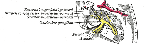
The Facial Nerve. Illustration showing the course and anatomical connections of the facial nerve within the temporal bone, with emphasis on the geniculate ganglion.
Henry Vandyke Carter, Public Domain, via Wikimedia Commons
(Click Image to Enlarge)
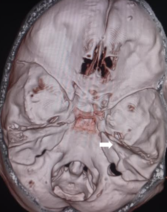
The Petrous Part of the Temporal Bone. The petrous part in the image is indicated by the white arrow. This arrow points to the petrous part of the temporal bone.
Contributed by S Munakomi, MD
(Click Image to Enlarge)
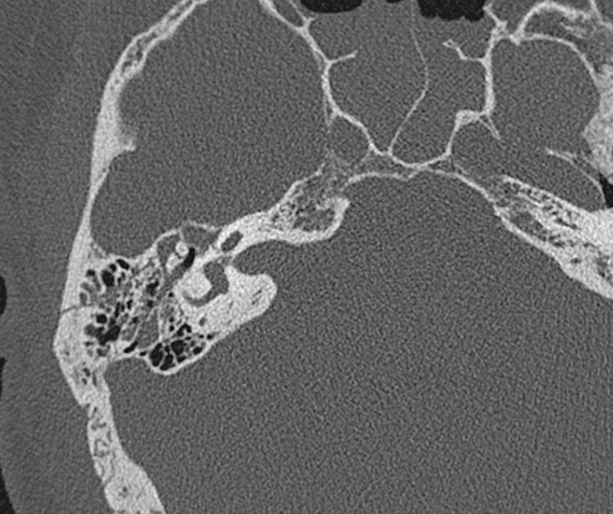
Right Temporal Bone Fracture Observed on CT Scan. Computed tomography (CT) scan demonstrates a fracture of the right temporal bone with ossicular disruption.
Contributed by S Lange, MD
(Click Image to Enlarge)
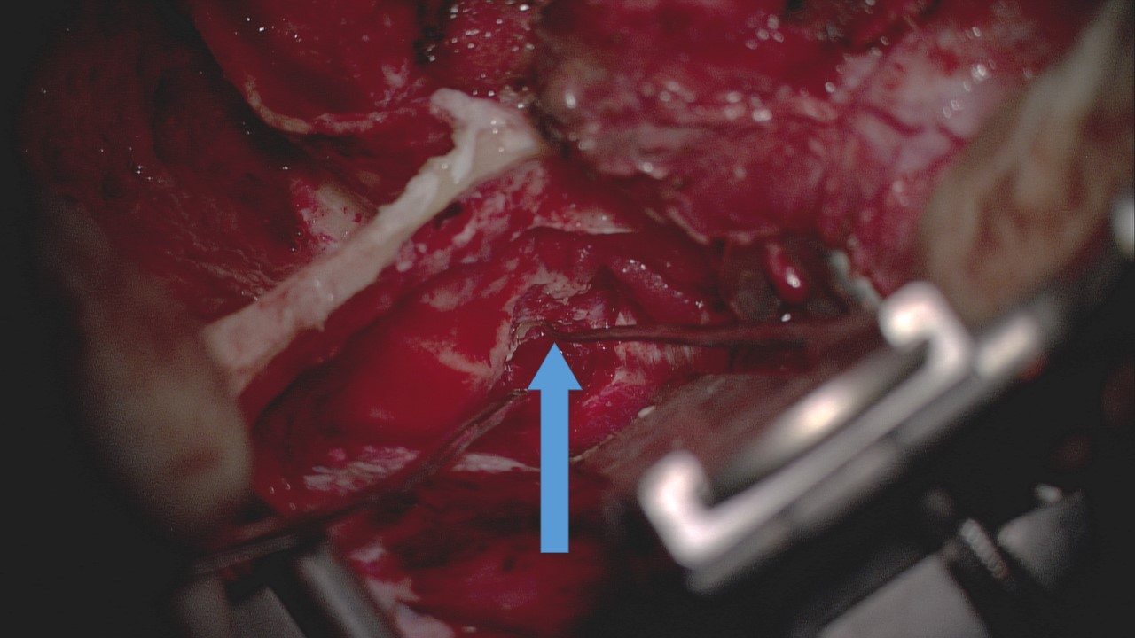
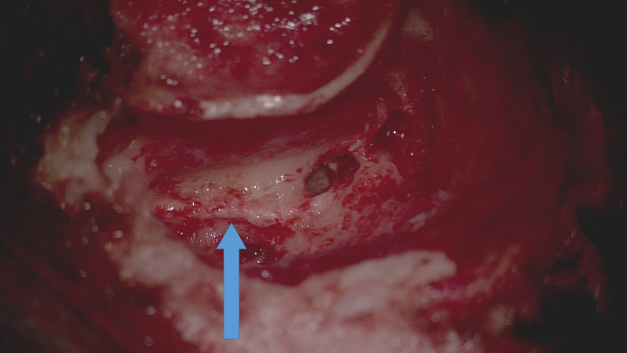
Middle Fossa Craniotomy Approach for Facial Nerve Decompression. The blue arrow indicates the approximate location of the facial nerve within the internal auditory canal during a middle fossa craniotomy approach.
Contributed by DS Ruhl, MD, MSPH, and AG Vincent, MD, FACS
(Click Image to Enlarge)
Transmastoid Approach to Facial Nerve Decompression. The blue arrow indicates the facial nerve during a transmastoid decompression procedure.
Contributed by DS Ruhl, MD, MSPH, and AG Vincent, MD, FACS
(Click Image to Enlarge)
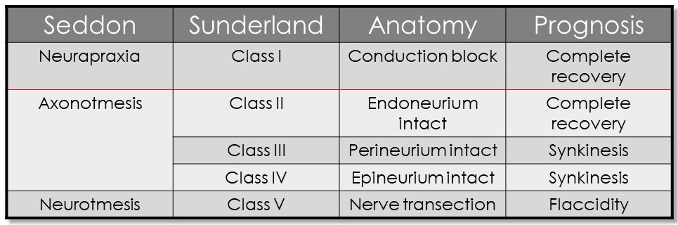
Seddon and Sunderland Classifications of Nerve Injury. This table presents the classifications of nerve injury according to Seddon and Sunderland, correlating each type with the microanatomy involved and the expected degree of recovery. Injuries below the red line are associated with Wallerian degeneration.
Contributed by MH Hohman, MD, FACS
(Click Image to Enlarge)
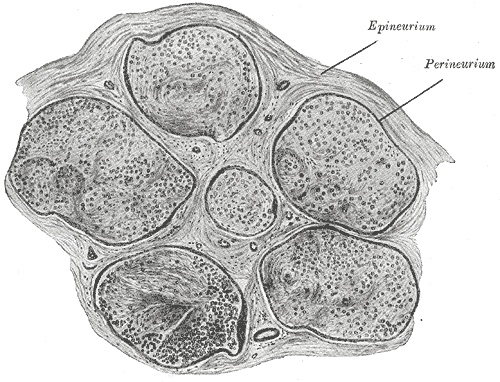
Neurology—Nerve Fascicle and Connective Tissue Layers. In the peripheral nervous system, nerve fibers are organized into fascicles, which are surrounded and supported by connective tissue layers, including the epineurium, perineurium, and endoneurium.
Henry Vandyke Carter, Public Domain, via Wikimedia Commons
References
Yalçıner G, Kutluhan A, Bozdemir K, Cetin H, Tarlak B, Bilgen AS. Temporal bone fractures: evaluation of 77 patients and a management algorithm. Ulusal travma ve acil cerrahi dergisi = Turkish journal of trauma & emergency surgery : TJTES. 2012 Sep:18(5):424-8. doi: 10.5505/tjtes.2012.98957. Epub [PubMed PMID: 23188604]
Level 2 (mid-level) evidenceCannon CR, Jahrsdoerfer RA. Temporal bone fractures. Review of 90 cases. Archives of otolaryngology (Chicago, Ill. : 1960). 1983 May:109(5):285-8 [PubMed PMID: 6847478]
Level 3 (low-level) evidenceBrodie HA, Thompson TC. Management of complications from 820 temporal bone fractures. The American journal of otology. 1997 Mar:18(2):188-97 [PubMed PMID: 9093676]
Level 2 (mid-level) evidenceYanagihara N, Murakami S, Nishihara S. Temporal bone fractures inducing facial nerve paralysis: a new classification and its clinical significance. Ear, nose, & throat journal. 1997 Feb:76(2):79-80, 83-6 [PubMed PMID: 9046695]
Hasso AN, Ledington JA. Traumatic injuries of the temporal bone. Otolaryngologic clinics of North America. 1988 May:21(2):295-316 [PubMed PMID: 3258654]
Wiet RJ, Valvassori GE, Kotsanis CA, Parahy C. Temporal bone fractures. State of the art review. The American journal of otology. 1985 May:6(3):207-15 [PubMed PMID: 4003527]
Level 3 (low-level) evidenceTravis LW, Stalnaker RL, Melvin JW. Impact trauma of the human temporal bone. The Journal of trauma. 1977 Oct:17(10):761-6 [PubMed PMID: 909117]
Johnson F, Semaan MT, Megerian CA. Temporal bone fracture: evaluation and management in the modern era. Otolaryngologic clinics of North America. 2008 Jun:41(3):597-618, x. doi: 10.1016/j.otc.2008.01.006. Epub [PubMed PMID: 18436001]
Ghorayeb BY, Yeakley JW. Temporal bone fractures: longitudinal or oblique? The case for oblique temporal bone fractures. The Laryngoscope. 1992 Feb:102(2):129-34 [PubMed PMID: 1738283]
Level 3 (low-level) evidenceDiaz RC, Cervenka B, Brodie HA. Treatment of Temporal Bone Fractures. Journal of neurological surgery. Part B, Skull base. 2016 Oct:77(5):419-29. doi: 10.1055/s-0036-1584197. Epub 2016 Jun 2 [PubMed PMID: 27648399]
Feng Y, Patel NS, Burrows AM, Lane JI, Raghunathan A, Van Gompel JJ, Carlson ML. Expansile Traumatic Neuroma of the Intratemporal Facial Nerve. Journal of neurological surgery reports. 2019 Jan:80(1):e10-e13. doi: 10.1055/s-0039-1685212. Epub 2019 Apr 1 [PubMed PMID: 30941279]
Gordin E, Lee TS, Ducic Y, Arnaoutakis D. Facial nerve trauma: evaluation and considerations in management. Craniomaxillofacial trauma & reconstruction. 2015 Mar:8(1):1-13. doi: 10.1055/s-0034-1372522. Epub [PubMed PMID: 25709748]
Vashishth A, Singh Nagar TR, Mandal S, Venkatachalam VP. Extensive intratemporal cholesteatomas: presentation, complications and surgical outcomes. European archives of oto-rhino-laryngology : official journal of the European Federation of Oto-Rhino-Laryngological Societies (EUFOS) : affiliated with the German Society for Oto-Rhino-Laryngology - Head and Neck Surgery. 2015 Feb:272(2):289-95. doi: 10.1007/s00405-013-2852-y. Epub 2013 Dec 8 [PubMed PMID: 24318471]
Level 2 (mid-level) evidenceNager GT, Proctor B. Anatomic variations and anomalies involving the facial canal. Otolaryngologic clinics of North America. 1991 Jun:24(3):531-53 [PubMed PMID: 1762775]
Vianna M, Adams M, Schachern P, Lazarini PR, Paparella MM, Cureoglu S. Differences in the diameter of facial nerve and facial canal in bell's palsy--a 3-dimensional temporal bone study. Otology & neurotology : official publication of the American Otological Society, American Neurotology Society [and] European Academy of Otology and Neurotology. 2014 Mar:35(3):514-8. doi: 10.1097/MAO.0000000000000240. Epub [PubMed PMID: 24518410]
Ottaiano AC, Gomez GD, Freddi TAL. The Facial Nerve: Anatomy and Pathology. Seminars in ultrasound, CT, and MR. 2023 Apr:44(2):71-80. doi: 10.1053/j.sult.2022.11.005. Epub 2022 Nov 28 [PubMed PMID: 37055142]
Guttal C, Prasad K, Kv V, G I, S G. Revisiting the Intratemporal Course of the Facial Nerve. Cureus. 2024 Nov:16(11):e73280. doi: 10.7759/cureus.73280. Epub 2024 Nov 8 [PubMed PMID: 39650983]
Remenschneider AK, Michalak S, Kozin ED, Barber S, De Venecia RK, Hadlock TA, Jung DH. Is Serial Electroneuronography Indicated Following Temporal Bone Trauma? Otology & neurotology : official publication of the American Otological Society, American Neurotology Society [and] European Academy of Otology and Neurotology. 2017 Apr:38(4):572-576. doi: 10.1097/MAO.0000000000001337. Epub [PubMed PMID: 28114180]
Darrouzet V, Duclos JY, Liguoro D, Truilhe Y, De Bonfils C, Bebear JP. Management of facial paralysis resulting from temporal bone fractures: Our experience in 115 cases. Otolaryngology--head and neck surgery : official journal of American Academy of Otolaryngology-Head and Neck Surgery. 2001 Jul:125(1):77-84 [PubMed PMID: 11458219]
Level 2 (mid-level) evidenceHouse JW, Brackmann DE. Facial nerve grading system. Otolaryngology--head and neck surgery : official journal of American Academy of Otolaryngology-Head and Neck Surgery. 1985 Apr:93(2):146-7 [PubMed PMID: 3921901]
Allen KP, Hatanpaa KJ, Lemeshev Y, Isaacson B, Kutz JW. Intratemporal traumatic neuromas of the facial nerve: evidence for multiple etiologies. Otology & neurotology : official publication of the American Otological Society, American Neurotology Society [and] European Academy of Otology and Neurotology. 2014 Feb:35(2):e69-72. doi: 10.1097/MAO.0000000000000136. Epub [PubMed PMID: 24270721]
Level 3 (low-level) evidenceNash JJ, Friedland DR, Boorsma KJ, Rhee JS. Management and outcomes of facial paralysis from intratemporal blunt trauma: a systematic review. The Laryngoscope. 2010:120 Suppl 4():S214. doi: 10.1002/lary.21681. Epub [PubMed PMID: 21225812]
Level 1 (high-level) evidenceKim J, Moon IS, Shim DB, Lee WS. The effect of surgical timing on functional outcomes of traumatic facial nerve paralysis. The Journal of trauma. 2010 Apr:68(4):924-9. doi: 10.1097/TA.0b013e3181a8b2d9. Epub [PubMed PMID: 20032793]
Level 2 (mid-level) evidenceGordon T. Peripheral Nerve Regeneration and Muscle Reinnervation. International journal of molecular sciences. 2020 Nov 17:21(22):. doi: 10.3390/ijms21228652. Epub 2020 Nov 17 [PubMed PMID: 33212795]
Gantz BJ, Rubinstein JT, Gidley P, Woodworth GG. Surgical management of Bell's palsy. The Laryngoscope. 1999 Aug:109(8):1177-88 [PubMed PMID: 10443817]
Prell J, Skinner S. EMG monitoring. Handbook of clinical neurology. 2022:186():67-81. doi: 10.1016/B978-0-12-819826-1.00002-8. Epub [PubMed PMID: 35772900]
Aslan H, Songu M, Eren E, Başoğlu MS, Özkul Y, Ateş D, Katilmiş H, Güvenç G. Results of decompression with middle cranial fossa approach or traumatic intratemporal fascial nerve injury. The Journal of craniofacial surgery. 2014 Jul:25(4):1305-8. doi: 10.1097/SCS.0000000000000772. Epub [PubMed PMID: 25006913]
Level 2 (mid-level) evidencePatel A, Groppo E. Management of temporal bone trauma. Craniomaxillofacial trauma & reconstruction. 2010 Jun:3(2):105-13. doi: 10.1055/s-0030-1254383. Epub [PubMed PMID: 22110824]
Wang Y, Cruz CD, Stern BJ. Approach to Facial Weakness. Seminars in neurology. 2021 Dec:41(6):673-685. doi: 10.1055/s-0041-1726358. Epub 2021 Nov 26 [PubMed PMID: 34826871]
Luu NN, Chorath KT, May BR, Bhuiyan N, Moreira AG, Rajasekaran K. Clinical practice guidelines in idiopathic facial paralysis: systematic review using the appraisal of guidelines for research and evaluation (AGREE II) instrument. Journal of neurology. 2021 May:268(5):1847-1856. doi: 10.1007/s00415-020-10345-0. Epub 2021 Jan 3 [PubMed PMID: 33389026]
Level 1 (high-level) evidenceHohman MH, Hadlock TA. Etiology, diagnosis, and management of facial palsy: 2000 patients at a facial nerve center. The Laryngoscope. 2014 Jul:124(7):E283-93. doi: 10.1002/lary.24542. Epub 2014 Jan 15 [PubMed PMID: 24431233]
Level 2 (mid-level) evidenceHohman MH, Bhama PK, Hadlock TA. Epidemiology of iatrogenic facial nerve injury: a decade of experience. The Laryngoscope. 2014 Jan:124(1):260-5. doi: 10.1002/lary.24117. Epub 2013 Apr 18 [PubMed PMID: 23606475]
Level 2 (mid-level) evidenceQuesnel AM, Lindsay RW, Hadlock TA. When the bell tolls on Bell's palsy: finding occult malignancy in acute-onset facial paralysis. American journal of otolaryngology. 2010 Sep-Oct:31(5):339-42. doi: 10.1016/j.amjoto.2009.04.003. Epub 2009 Jun 24 [PubMed PMID: 20015776]
Level 3 (low-level) evidenceMinor LB, Solomon D, Zinreich JS, Zee DS. Sound- and/or pressure-induced vertigo due to bone dehiscence of the superior semicircular canal. Archives of otolaryngology--head & neck surgery. 1998 Mar:124(3):249-58 [PubMed PMID: 9525507]
Level 3 (low-level) evidenceKang HM, Kim MG, Boo SH, Kim KH, Yeo EK, Lee SK, Yeo SG. Comparison of the clinical relevance of traditional and new classification systems of temporal bone fractures. European archives of oto-rhino-laryngology : official journal of the European Federation of Oto-Rhino-Laryngological Societies (EUFOS) : affiliated with the German Society for Oto-Rhino-Laryngology - Head and Neck Surgery. 2012 Aug:269(8):1893-9. doi: 10.1007/s00405-011-1849-7. Epub 2011 Nov 26 [PubMed PMID: 22120750]
Level 2 (mid-level) evidenceRafferty MA, Mc Conn Walsh R, Walsh MA. A comparison of temporal bone fracture classification systems. Clinical otolaryngology : official journal of ENT-UK ; official journal of Netherlands Society for Oto-Rhino-Laryngology & Cervico-Facial Surgery. 2006 Aug:31(4):287-91 [PubMed PMID: 16911644]
Level 2 (mid-level) evidencePourmomeny AA, Asadi S. Management of synkinesis and asymmetry in facial nerve palsy: a review article. Iranian journal of otorhinolaryngology. 2014 Oct:26(77):251-6 [PubMed PMID: 25320703]
Robinson MW, Baiungo J. Facial Rehabilitation: Evaluation and Treatment Strategies for the Patient with Facial Palsy. Otolaryngologic clinics of North America. 2018 Dec:51(6):1151-1167. doi: 10.1016/j.otc.2018.07.011. Epub 2018 Sep 24 [PubMed PMID: 30262166]
Miller MQ, Hadlock TA. Beyond Botox: Contemporary Management of Nonflaccid Facial Palsy. Facial plastic surgery & aesthetic medicine. 2020 Mar/Apr:22(2):65-70. doi: 10.1089/fpsam.2020.0009. Epub [PubMed PMID: 32130060]
Hohman MH, Lee LN, Hadlock TA. Two-step highly selective neurectomy for refractory periocular synkinesis. The Laryngoscope. 2013 Jun:123(6):1385-8. doi: 10.1002/lary.23873. Epub 2013 Jan 11 [PubMed PMID: 23315713]
Level 2 (mid-level) evidenceVincent AG, Bevans SE, Robitschek JM, Wind GG, Hohman MH. Masseteric-to-Facial Nerve Transfer and Selective Neurectomy for Rehabilitation of the Synkinetic Smile. JAMA facial plastic surgery. 2019 Dec 1:21(6):504-510. doi: 10.1001/jamafacial.2019.0689. Epub [PubMed PMID: 31465094]
Silver AL, Lindsay RW, Cheney ML, Hadlock TA. Thin-profile platinum eyelid weighting: a superior option in the paralyzed eye. Plastic and reconstructive surgery. 2009 Jun:123(6):1697-1703. doi: 10.1097/PRS.0b013e3181a65a56. Epub [PubMed PMID: 19483568]
Klebuc MJA. Facial reanimation using the masseter-to-facial nerve transfer. Plastic and reconstructive surgery. 2011 May:127(5):1909-1915. doi: 10.1097/PRS.0b013e31820e9138. Epub [PubMed PMID: 21532419]
Level 2 (mid-level) evidenceKoo WY, Park SO, Ahn HC, Ryu SR. Facial reanimation using the hypoglossal nerve and ansa cervicalis: a short-term retrospective analysis of surgical outcomes. Archives of craniofacial surgery. 2021 Dec:22(6):303-309. doi: 10.7181/acfs.2021.00444. Epub 2021 Dec 20 [PubMed PMID: 34974685]
Level 2 (mid-level) evidenceLabbé D. [Lengthening temporalis myoplasty]. Revue de stomatologie et de chirurgie maxillo-faciale. 2002 Apr:103(2):79-83 [PubMed PMID: 11997734]
Harii K, Ohmori K, Torii S. Free gracilis muscle transplantation, with microneurovascular anastomoses for the treatment of facial paralysis. A preliminary report. Plastic and reconstructive surgery. 1976 Feb:57(2):133-43 [PubMed PMID: 1250883]
Level 3 (low-level) evidenceKleiss IJ, Hohman MH, Susarla SM, Marres HA, Hadlock TA. Health-related quality of life in 794 patients with a peripheral facial palsy using the FaCE Scale: a retrospective cohort study. Clinical otolaryngology : official journal of ENT-UK ; official journal of Netherlands Society for Oto-Rhino-Laryngology & Cervico-Facial Surgery. 2015 Dec:40(6):651-6. doi: 10.1111/coa.12434. Epub [PubMed PMID: 25858429]
Level 2 (mid-level) evidenceRatilal BO, Costa J, Sampaio C, Pappamikail L. Antibiotic prophylaxis for preventing meningitis in patients with basilar skull fractures. The Cochrane database of systematic reviews. 2011 Aug 10:(8):CD004884. doi: 10.1002/14651858.CD004884.pub3. Epub 2011 Aug 10 [PubMed PMID: 21833952]
Level 1 (high-level) evidenceHadlock TA, Greenfield LJ, Wernick-Robinson M, Cheney ML. Multimodality approach to management of the paralyzed face. The Laryngoscope. 2006 Aug:116(8):1385-9 [PubMed PMID: 16885741]
Level 2 (mid-level) evidenceBhama PK, Weinberg JS, Lindsay RW, Hohman MH, Cheney ML, Hadlock TA. Objective outcomes analysis following microvascular gracilis transfer for facial reanimation: a review of 10 years' experience. JAMA facial plastic surgery. 2014 Mar-Apr:16(2):85-92. doi: 10.1001/jamafacial.2013.2463. Epub [PubMed PMID: 24481538]
Level 2 (mid-level) evidenceVincent AG, Bevans SE, Robitschek JM, Groom KL, Herr MW, Hohman MH. Sterno-omohyoid Free Flap for Dual-Vector Dynamic Facial Reanimation. The Annals of otology, rhinology, and laryngology. 2020 Feb:129(2):195-200. doi: 10.1177/0003489419875473. Epub 2019 Oct 3 [PubMed PMID: 31578078]
Sun GH, Shoman NM, Samy RN, Cornelius RS, Koch BL, Pensak ML. Do contemporary temporal bone fracture classification systems reflect concurrent intracranial and cervical spine injuries? The Laryngoscope. 2011 May:121(5):929-32. doi: 10.1002/lary.21718. Epub [PubMed PMID: 21520104]
Level 2 (mid-level) evidenceAlfrey EJ, Tracy M, Alfrey JR, Carroll M, Aranda-Wikman ED, Arora T, Maa J, Minnis J. Helmet Usage Reduces Serious Head Injury Without Decreasing Concussion After Bicycle Riders Crash. The Journal of surgical research. 2021 Jan:257():593-596. doi: 10.1016/j.jss.2020.08.009. Epub 2020 Sep 12 [PubMed PMID: 32932191]
Rughani AI, Lin CT, Ares WJ, Cushing DA, Horgan MA, Tranmer BI, Jewell RP, Florman JE. Helmet use and reduction in skull fractures in skiers and snowboarders admitted to the hospital. Journal of neurosurgery. Pediatrics. 2011 Mar:7(3):268-71. doi: 10.3171/2010.12.PEDS10415. Epub [PubMed PMID: 21361765]
Level 2 (mid-level) evidence