Introduction
Diabetes mellitus is a metabolic disorder characterized by chronic hyperglycemia. About 589 million adults aged 20 to 79 years are living with diabetes globally as of 2025, and diabetes-related deaths are estimated at approximately 3 to 4 million annually.[1] (Source: International Diabetes Foundation, 2025) Individuals with diabetes are at increased risk of pedal ulceration due to microvascular, neuropathic, and biomechanical alterations in the foot. Neuropathy reduces pedal sensation, predisposing the foot to pressure- and trauma-related injuries. Microvascular dysfunction impairs blood flow to the lower extremities, delaying wound healing.
Hemoglobin A1c (HbA1c) indicates mean blood glucose over 2 to 3 months. Both the American Diabetes Association (ADA) and the International Expert Committee recommend an HbA1c of 6.5% or greater for diabetes diagnosis.[2] Each 1% increase in HbA1c increases peripheral vascular disease risk by 25% to 28%.[3]
Patients with diabetes face an elevated risk of lower extremity amputation, increased healthcare costs, and reduced quality of life. In a systematic review of nontraumatic amputations among patients with diabetes and peripheral vascular disease, the 5-year mortality rate after below-the-knee amputation was 40% to 82%, and 40% to 90% following above-the-knee amputation.[4] Many complications are preventable through annual foot examination and routine patient-performed foot care. This activity delves into the anatomy, indications, equipment, personnel, preparation, technique, complications, and clinical significance of diabetic foot care.
Anatomy and Physiology
Register For Free And Read The Full Article
 Search engine and full access to all medical articles
- 10 free questions in your specialty
- Free CME/CE Activities
- Free daily question in your email
- Save favorite articles to your dashboard
- Emails offering discounts
Learn more about a Subscription to StatPearls Point-of-Care
Anatomy and Physiology
The diabetic foot exhibits multiple structural and functional changes compared to a nondiabetic foot. These modifications encompass musculoskeletal, dermatologic, vascular, and neurological adaptations.
Musculoskeletal Manifestations
Musculoskeletal changes include atrophy of intrinsic musculature, limited joint mobility, alterations in foot type, and the development of ankle equinus. Limited dorsiflexion of the 1st metatarsophalangeal joint (hallux limitus) is frequently observed in patients with diabetes and is often associated with thickening of the Achilles tendon and plantar fascia. This tendon and fascia thickening contributes to increased foot rigidity, greater pes planus, and potential gait instability.
Longstanding diabetes leads to disorganization of the pedal musculature and infiltration with adipose tissue. Atrophy of intrinsic foot muscles relative to extrinsic muscles contributes to deformities, including hammertoes and claw toe deformities.[5]
Hammertoes and claw toes, in combination with hallux limitus, are associated with an elevated risk of ulcer formation. Bus et al demonstrated that plantar pressures at the metatarsal heads increase significantly with the severity of toe deformities. The study further indicated a distal-to-proximal load transfer in affected toes, potentially exacerbated by displacement of the distal fat pad.[6]
Ankle equinus, defined as dorsiflexion less than 5°, is prevalent in patients with diabetes. Searle et al reported that this limitation correlates with increased forefoot pressure and heightened risk of tissue breakdown, both during barefoot ambulation and while wearing shoes.[7]
Dermatologic Changes
Dermatologic integrity is a critical function of human skin. However, chronic diabetes induces multiple alterations that compromise this protective barrier. Autonomic dysfunction associated with diabetes reduces perspiration in the foot, predisposing to fissuring and xerosis.[8]
Repetitive mechanical stress and localized pressure, combined with neuropathy, promote inflammation and ulcer formation. Plantar skin thickness is reduced in patients with type 2 diabetes and neuropathy compared to patients with diabetes without neuropathy, further increasing the risk of ulceration (see Image. Anatomical Model of a Diabetic Foot).[9] Perilesional tissue remains vulnerable to rapid breakdown and recurrent ulceration even after healing.[10]
Vascular Abnormalities
Diabetes also impairs lower extremity perfusion, contributing to delayed wound healing and increased susceptibility to tissue injury. Three primary arteries and their branches perfuse the 6 angiosomes of the foot and ankle. The posterior tibial artery, a branch of the popliteal artery, supplies the plantar surface of the foot. The fibular artery arises from the posterior tibial artery and perfuses the anterolateral ankle and rearfoot. The anterior tibial artery originates from the popliteal artery and continues distally as the dorsalis pedis artery, supplying the anterior ankle and dorsal foot.[11]
Individuals with diabetes are at increased risk of peripheral arterial disease (PAD), characterized by atherosclerotic obstruction of arteries supplying the lower extremities. Over 50% of affected individuals remain asymptomatic. However, some experience intermittent claudication—exercise-induced lower extremity pain relieved by rest—or rest pain, and in severe cases, tissue loss and gangrene.
Neurological Alterations
The foot receives innervation from 5 principal nerves—tibial, superficial fibular, deep fibular, sural, and saphenous—and their branches. The tibial nerve, a branch of the sciatic nerve, divides into the medial and lateral plantar nerves, which further give rise to the digital nerves. The tibial nerve provides motor innervation to the posterior compartment muscles of the lower leg and sensory innervation to the plantar foot and heel. The superficial fibular nerve originates from the common fibular nerve and branches into the medial and intermediate dorsal cutaneous nerves, innervating the fibularis longus and brevis muscles and providing sensory input to the anterior lower leg and dorsal foot and toes, excluding the 1st webspace.
The deep fibular nerve, also a branch of the common fibular nerve, supplies motor fibers to the anterior compartment muscles and sensory fibers to the 1st webspace. The sural nerve arises from contributions of the tibial and common fibular nerves and provides sensory innervation to the posterolateral lower leg and foot. The saphenous nerve, a branch of the femoral nerve, supplies sensory innervation to the medial distal leg, ankle, and foot.[12][13]
Diabetic neuropathy is a common neurological complication of diabetes, with up to 50% of cases remaining asymptomatic.[14][15] Distal symmetrical polyneuropathy is the most prevalent form and may involve sensory, motor, or mixed nerve fiber dysfunction. Large fiber neuropathy (Aα/β fiber damage) produces painless paresthesia, diminished vibration sense, impaired joint position sense, reduced touch and pressure sensation, and absent or decreased ankle reflexes. Small fiber neuropathy (myelinated Aδ and unmyelinated C fiber damage) manifests as painful, burning sensations with impaired pain and temperature perception.
Peripheral neuropathy typically begins in the distal toes and progresses proximally. In advanced stages, a stocking-glove sensory distribution may develop in the upper extremities. Nocturnal worsening of symptoms is common, and motor involvement can lead to muscle weakness. The pathogenesis involves multiple mechanisms, including chronic hyperglycemia, metabolic alterations, direct axonal injury, and nerve ischemia. However, the precise contribution of each mechanism remains under investigation.[16][17]
A dreaded complication of uncontrolled diabetes and peripheral neuropathy is Charcot neuroarthropathy. This condition likely results from both neurovascular changes, including arteriovenous shunting that increases blood flow and bone resorption, and repetitive microtrauma.[18] These processes lead to joint collapse and severe pedal deformities. The tarsometatarsal joint is most commonly affected, resulting in a rocker-bottom deformity.
Patients with Charcot neuroarthropathy have a 17% annual risk of developing ulceration. The risk of lower extremity amputation in those with ulceration is 12 times higher compared to patients with Charcot neuroarthropathy without ulceration.[19] Early recognition and management improve outcomes. Clinicians should suspect Charcot neuroarthropathy when a patient with diabetes presents with a warm, erythematous, edematous foot and structural abnormalities.
Indications
All patients with diabetes should receive structured education on proper diabetic foot care. Prevention of diabetic foot complications involves identifying at-risk feet, performing daily examination and inspection, providing education to patients, families, and healthcare providers, recommending appropriate shoegear, and initiating timely treatment of preulcerative lesions. Patients at higher risk must be referred to podiatry for specialized management and ongoing monitoring. The ADA recommends the following diabetic foot screening by a qualified medical professional:
- A thorough foot assessment must be conducted at least once per year to identify factors that increase the risk of ulceration or amputation. The evaluation should include inspection of the skin, identification of structural deformities, neurological testing, and vascular assessment, including palpation of leg and foot pulses.
- Patients with sensory deficits, previous ulceration, or a history of amputation should have their feet examined at every clinical encounter.
- Prior occurrences of ulceration, amputation, Charcot foot, revascularization procedures, cigarette use, retinopathy, and kidney disease must be documented. Current neuropathic symptoms (pain, burning, numbness) and vascular manifestations (leg fatigue, claudication) should be evaluated.
- PAD screening must be performed by assessing lower-extremity pulses, capillary refill, dependent rubor, pallor on elevation, and venous filling time.
- Patients reporting leg fatigue, claudication, rest pain relieved by dependency, or exhibiting diminished or absent pedal pulses should be referred for ankle-brachial index (ABI) with toe pressures and additional vascular evaluation as indicated.
- A podiatrist-led interprofessional team, including other relevant specialists, should manage patients with foot ulcers or high-risk feet (eg, those on dialysis, with Charcot foot, prior ulcers or amputation, or PAD).
- Patients who smoke and have prior lower-extremity complications, loss of protective sensation, structural deformities, or PAD should be given a referral to a foot care specialist for continuous preventive treatment and lifelong monitoring. These patients should also receive education on the benefits of smoking cessation and be referred for counseling as appropriate.
- All patients with diabetes, including those with sensory loss, should receive education on proper foot self-care, including daily inspection and palpation or visual examination using an unbreakable mirror to detect early problems.
- Therapeutic footwear designed for high-risk patients is recommended for individuals with diabetes who have loss of protective sensation, foot deformities, current or prior ulcers, callus formation, poor circulation, or a history of amputation.
Patient education is a mainstay of diabetic foot prevention, enabling daily self-monitoring and early recognition of complications. Ongoing instruction and reinforcement by healthcare teams can substantially reduce morbidity and preserve limb function.
Contraindications
Proper diabetic foot care has no contraindications. Preventive foot care is appropriate for every patient with diabetes and poses minimal risk.
Equipment
A basic diabetic foot examination requires minimal specialized instruments and may be performed by most qualified physicians, including primary care clinicians and podiatrists. Neurological assessment typically includes a Semmes-Weinstein monofilament test (SWMT) for evaluating neuropathy and protective sensation. A 128-Hz tuning fork may be used to assess vibratory sensation, and cotton wool can evaluate tactile sensation. Vascular examination may involve Doppler ultrasonography to assess lower-extremity blood flow.
Caregivers may encounter ulcerations during diabetic foot care. If present and requiring debridement or offloading, instruments such as scalpels, tissue nippers, and offloading padding (eg, felt, foam, or cushion pads) should be available. For advanced wound care, a comprehensive set of products should be accessible, including gauze, cleansing solutions (eg, saline, hydrogen peroxide, acetic acid), topical antimicrobials (eg, povidone-iodine, cadexomer iodine, silver, medical-grade honey), moisture-retentive dressings (eg, films, foams, alginates, hydrogels, hydrocolloids), vacuum-assisted closure devices, and bioengineered dressings.[20]
Personnel
An interprofessional approach to managing the diabetic foot and its potential complications can reduce amputation rates by up to 85%. Key team members include endocrinology, diabetology, vascular surgery, podiatry, orthotics, and prosthetics specialists, wound care nurses, and patient educators. Depending on individual medical conditions and infection status, additional specialties, such as infectious disease, nephrology, cardiology, and dermatology, may also contribute. The primary objectives of interprofessional care are to medically optimize comorbidities, prevent foot complications, and manage existing pedal pathology.
Preparation
Patients with diabetes should remove both shoes and socks prior to examination. Feet should be thoroughly cleaned and dried before any foot assessment or dressing change. Caregivers should wash their hands before and after performing a dressing change for a diabetic foot ulcer. A clean and sanitary environment should be maintained to reduce the likelihood of contamination and infection.
Technique or Treatment
The clinical diabetic foot examination comprises 4 major components: dermatological, vascular, neurological, and musculoskeletal. Each component is explained in detail below.
Dermatological Assessment
The dermatological examination of the diabetic foot should encompass a comprehensive evaluation of the entire foot and ankle, including interdigital spaces and nails. Hyperkeratotic areas should be debrided. Preulcerative and ulcerative lesions require detailed documentation of location, size, depth, wound base characteristics, periwound skin, undermining, sinus tracts, probing to bone, exudate quality, and signs of infection, such as erythema, warmth, malodor, or crepitus.
A temperature difference exceeding 3 °C to 4 °C compared to the contralateral foot may indicate infection or acute Charcot neuroarthropathy. Additionally, a prior history of ulceration, treatments, and preventive interventions should be recorded. Baseline radiographs are recommended for new ulcerations, and serial imaging should be obtained when osteomyelitis is suspected to monitor progression and guide management (see Images. Right Foot Osteomyelitis on Magnetic Resonance Imaging; Right Foot Radiograph After Partial 5th Ray Resection).[21]
Vascular Evaluation
Clinical evaluation of vascularity and PAD begins with a detailed medical history, including PAD risk factors, claudication, rest pain, and prior nonhealing wounds. Dorsalis pedis and posterior tibial artery palpation forms the baseline of diabetic foot exams, while assessment of popliteal and femoral pulses further delineates PAD severity.
A decrease in pedal pulses may prompt evaluation with Doppler ultrasound or additional noninvasive PAD testing. Doppler ultrasound assessment of pedal arteries can help characterize blood flow and detect the severity of PAD. Flow patterns are classified as follows:
- Triphasic: normal
- Biphasic: mild arterial disease
- Monophasic: PAD with some risk of limb ischemia
- Absent: severe PAD, high risk of ischemia and limb loss
Noninvasive vascular tests include ABI, segmental pressures, pulse volume recording, toe pressures, transcutaneous oxygen pressure, and treadmill functional testing. The ADA recommends a screening ABI for individuals with diabetes older than 50 years, repeating every 5 years if normal. A screening ABI should be considered in patients younger than 50 with additional PAD risk factors (eg, smoking, hypertension, hyperlipidemia, diabetes duration >10 years). Any findings suggestive of PAD warrant referral to vascular surgery.[22]
Neurological Examination
The neurological examination of the diabetic foot includes assessment of the Achilles reflex, evaluation using the SWMT, vibratory sensation testing with a 128-Hz tuning fork, and pinprick sensory testing. The Achilles reflex should be assessed on both legs using a reflex hammer. Reflex responses are graded as follows:
- 0+: absent
- 1+: decreased
- 2+: brisk, normal
- 3+: increased
- 4+: increased with clonus
The 5.07/10 g SWMT is commonly used to assess pressure and light-touch sensation. Testing should include the following 10 sites on each foot:
- Distal 1st, 3rd, and 5th toes
- Plantar 1st, 3rd, and 5th metatarsal heads
- Plantar medial and lateral arches
- Plantar heel
- Dorsal 1st interspace
The 128-Hz nongraduated standard tuning fork is used to assess vibratory sensation. An abnormal finding may indicate diabetic neuropathy if the patient cannot perceive vibration. Pinprick testing, performed with a sharp pin device, evaluates the patient’s ability to differentiate between sharp and dull stimuli.[23][24][25]
Musculoskeletal Assessment
Musculoskeletal evaluation should assess lower extremity muscle strength, foot and ankle deformities, and joint range of motion, including flexibility and rigidity, to guide treatment planning. Patients with diabetes face a greater risk of pedal deformities, such as digital contractures and ankle equinus, which elevate the risk of ulceration. For instance, a flexible ankle equinus caused by gastrocnemius tightness can increase plantar foot pressures, leading to tissue breakdown in neuropathic patients. Individuals with pedal deformities should be referred to podiatry or appropriate surgical specialists for further management, as they may benefit from corrective procedures. For example, a digital flexor tenotomy can help prevent distal toe ulceration when tissue compromise is present.[26]
Patient and family education on proper diabetic foot care should be provided and reinforced during each visit.[27] Patients must understand the importance of wearing protective shoes indoors and outdoors, ensuring that footwear fits properly to prevent ulcers. Some patients may require specialized shoegear, which should be evaluated during the diabetic foot examination. Selection of appropriate offloading or protective modalities should be individualized based on biomechanical alterations, pressure points, and existing pathology. Options may include shoe modifications, temporary shoegear, toe spacers, orthoses, and offloading devices such as felt pads and foam to help protect the diabetic foot.[28]
Complications
Inadequate diabetic foot care significantly increases the risk of ulceration, infection, and limb loss. Armstrong and Harkless demonstrated that noncompliant patients, specifically those who missed more than 50% of scheduled appointments within 1 year, are 54 times more likely to develop pedal ulcerations and 20 times more likely to undergo amputation compared to compliant patients.[29] One study reported that individuals with diabetes fear major lower-extremity amputation more than death, foot infection, or end-stage renal disease.[30] Another study found that amputation exerts the largest detrimental effect on quality of life compared to other diabetes-related complications, including stroke, blindness, renal failure, heart failure, and myocardial infarction.[31] Consequently, high-risk limbs require intensive monitoring and management by specialists, such as podiatrists.
Clinical Significance
Proper diabetic foot care is a critical component of diabetes management for limb preservation. Foot ulceration is one of the leading causes of hospitalization and amputation among patients with diabetes. Most diabetic foot complications arise from ischemia, neuropathy, and infection. Patients with diabetes and neuropathy have a 7% to 10% annual risk of developing foot ulceration, which increases to 25% to 30% in individuals with additional risk factors, such as PAD, pedal deformities, or a history of ulceration or amputation. Approximately 85% of diabetes-related lower extremity amputations result from complications of foot ulceration. Risk of amputation rises with age. Patients older than 45 have an 8-fold increased risk of amputation, those older than 65 years a 12-fold risk, and individuals aged 65 to 74 years a 23-fold risk.
Enhancing Healthcare Team Outcomes
Diabetic foot care requires interprofessional collaboration. Patients receiving team-based care demonstrate reduced severity of amputation, shorter hospital stays, lower mortality rates, improved ulcer healing, and enhanced quality of life.[32]
Effective diabetes management is integral to foot care. Beyond routine endocrinology or diabetology oversight, group-based self-management programs improve body weight, fasting blood glucose, waist circumference, diabetic knowledge, and triglyceride levels. Monitoring responsibilities are shared among clinicians, nurses, and pharmacists, with results systematically relayed to the interprofessional team. Interprofessional interventions incorporating peer support have demonstrated superior reductions in HbA1c compared to peer-led groups.[33] Foot care nurses frequently perform assessments and provide patient education, but all team members share responsibility for training, reinforcing self-care, and preventing complications.
Nursing, Allied Health, and Interprofessional Team Interventions
Intensive patient education by the nursing staff, encompassing lifestyle management, podiatric care, routine foot examinations, and callus management, reduces the incidence of diabetic foot ulcerations and lowers amputation rates in high-risk patients. Additionally, such education contributes to improved metabolic parameters, including reductions in plasma glucose, blood pressure, and high-density lipoprotein cholesterol levels.[34]
Nursing, Allied Health, and Interprofessional Team Monitoring
Care of the diabetic foot is an interprofessional responsibility. Holstein et al reported a 75% reduction in major limb amputations in patients with diabetes following the establishment of an interprofessional foot clinic with increased revascularization rates.[35] Primary care physicians and nursing staff play a key role in identifying at-risk feet and providing education on proper care and monitoring. Individuals with elevated risk factors or pedal deformities should receive regular follow-up with podiatry. Development of ulcerations necessitates more frequent surveillance and may require referral to additional specialists, including vascular surgery, infectious disease, plastic surgery, and prosthetics.
Media
(Click Image to Enlarge)

Anatomical Model of a Diabetic Foot. This image shows a plastic anatomical model of the plantar surface of a foot. The model displays a deep, full-thickness ulceration on the 1st metatarsal head. A preulcerative lesion is also visible on the 5th metatarsal head.
Contributed by Aaron R. Chambers, DPM, FACFAS
(Click Image to Enlarge)
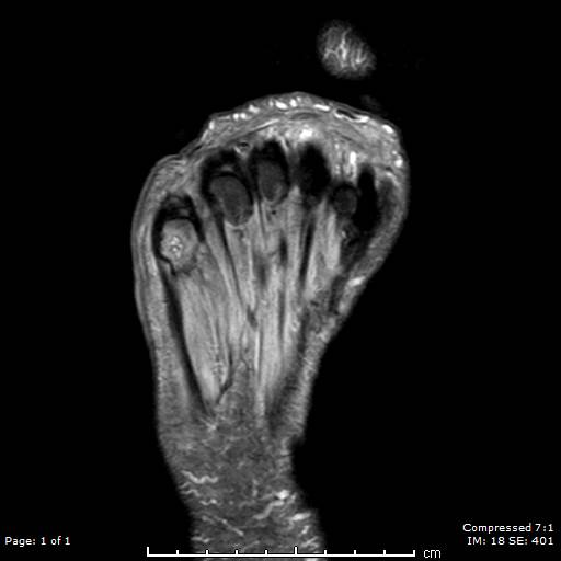
Right Foot Osteomyelitis on Magnetic Resonance Imaging. This T2-weighted image shows a patient’s right foot. The patient had a history of uncontrolled diabetes, peripheral neuropathy, and a longstanding ulceration under the 5th metatarsal head, resulting in osteomyelitis of the 5th metatarsal head and shaft. The patient subsequently underwent a partial 5th ray resection with long-term intravenous antibiotics for treatment and healed uneventfully.
Contributed by Aaron R. Chambers, DPM, FACFAS
(Click Image to Enlarge)
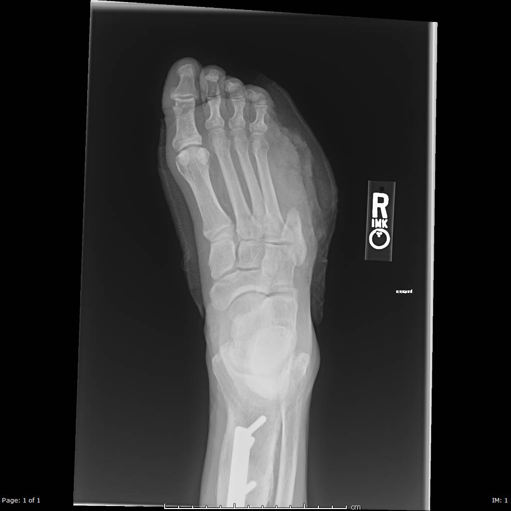
Right Foot Radiograph After Partial 5th Ray Resection. This x-ray shows a patient's right foot after undergoing a partial resection of the 5th metatarsal for acute osteomyelitis. The patient had a history of uncontrolled diabetes, peripheral neuropathy, and a chronic ulcer beneath the 5th metatarsal head, which led to osteomyelitis of the 5th metatarsal head and shaft. The patient underwent partial 5th ray resection followed by long-term intravenous antibiotic treatment and healed uneventfully.
Contributed by Aaron R. Chambers, DPM, FACFAS
References
Hossain MJ, Al-Mamun M, Islam MR. Diabetes mellitus, the fastest growing global public health concern: Early detection should be focused. Health science reports. 2024 Mar:7(3):e2004. doi: 10.1002/hsr2.2004. Epub 2024 Mar 22 [PubMed PMID: 38524769]
American Diabetes Association. Diagnosis and classification of diabetes mellitus. Diabetes care. 2011 Jan:34 Suppl 1(Suppl 1):S62-9. doi: 10.2337/dc11-S062. Epub [PubMed PMID: 21193628]
Lepäntalo M, Apelqvist J, Setacci C, Ricco JB, de Donato G, Becker F, Robert-Ebadi H, Cao P, Eckstein HH, De Rango P, Diehm N, Schmidli J, Teraa M, Moll FL, Dick F, Davies AH. Chapter V: Diabetic foot. European journal of vascular and endovascular surgery : the official journal of the European Society for Vascular Surgery. 2011 Dec:42 Suppl 2():S60-74. doi: 10.1016/S1078-5884(11)60012-9. Epub [PubMed PMID: 22172474]
Thorud JC, Plemmons B, Buckley CJ, Shibuya N, Jupiter DC. Mortality After Nontraumatic Major Amputation Among Patients With Diabetes and Peripheral Vascular Disease: A Systematic Review. The Journal of foot and ankle surgery : official publication of the American College of Foot and Ankle Surgeons. 2016 May-Jun:55(3):591-9. doi: 10.1053/j.jfas.2016.01.012. Epub 2016 Feb 19 [PubMed PMID: 26898398]
Level 1 (high-level) evidenceAllan J, Munro W, Figgins E. Foot deformities within the diabetic foot and their influence on biomechanics: A review of the literature. Prosthetics and orthotics international. 2016 Apr:40(2):182-92. doi: 10.1177/0309364615592705. Epub 2015 Jul 24 [PubMed PMID: 26209425]
Bus SA, Maas M, de Lange A, Michels RP, Levi M. Elevated plantar pressures in neuropathic diabetic patients with claw/hammer toe deformity. Journal of biomechanics. 2005 Sep:38(9):1918-25 [PubMed PMID: 16023481]
Level 1 (high-level) evidenceSearle A, Spink MJ, Chuter VH. Prevalence of ankle equinus and correlation with foot plantar pressures in people with diabetes. Clinical biomechanics (Bristol, Avon). 2018 Dec:60():39-44. doi: 10.1016/j.clinbiomech.2018.10.006. Epub 2018 Oct 5 [PubMed PMID: 30312937]
Markendeya N, Martina V, Mathew A, Srinivas CR. Sweat function in the diabetic foot. Indian journal of dermatology, venereology and leprology. 2004 Jan-Feb:70(1):18-9 [PubMed PMID: 17642551]
Kumar CG, Rajagopal KV, Hande HM, Maiya AG, Mayya SS. Intrinsic foot muscle and plantar tissue changes in type 2 diabetes mellitus. Journal of diabetes. 2015 Nov:7(6):850-7. doi: 10.1111/1753-0407.12254. Epub 2015 Mar 24 [PubMed PMID: 25496489]
Level 2 (mid-level) evidenceBrand PW. Tenderizing the foot. Foot & ankle international. 2003 Jun:24(6):457-61 [PubMed PMID: 12854665]
Level 3 (low-level) evidenceAttinger CE, Evans KK, Bulan E, Blume P, Cooper P. Angiosomes of the foot and ankle and clinical implications for limb salvage: reconstruction, incisions, and revascularization. Plastic and reconstructive surgery. 2006 Jun:117(7 Suppl):261S-293S [PubMed PMID: 16799395]
De Maeseneer M, Madani H, Lenchik L, Kalume Brigido M, Shahabpour M, Marcelis S, de Mey J, Scafoglieri A. Normal Anatomy and Compression Areas of Nerves of the Foot and Ankle: US and MR Imaging with Anatomic Correlation. Radiographics : a review publication of the Radiological Society of North America, Inc. 2015 Sep-Oct:35(5):1469-82. doi: 10.1148/rg.2015150028. Epub 2015 Aug 18 [PubMed PMID: 26284303]
Eglitis N, Horn JL, Benninger B, Nelsen S. The Importance of the Saphenous Nerve in Ankle Surgery. Anesthesia and analgesia. 2016 May:122(5):1704-6. doi: 10.1213/ANE.0000000000001168. Epub [PubMed PMID: 26859876]
American Diabetes Association Professional Practice Committee. 12. Retinopathy, Neuropathy, and Foot Care: Standards of Care in Diabetes-2025. Diabetes care. 2025 Jan 1:48(1 Suppl 1):S252-S265. doi: 10.2337/dc25-S012. Epub [PubMed PMID: 39651973]
American Diabetes Association. 10. Microvascular Complications and Foot Care: Standards of Medical Care in Diabetes-2018. Diabetes care. 2018 Jan:41(Suppl 1):S105-S118. doi: 10.2337/dc18-S010. Epub [PubMed PMID: 29222381]
Singh R, Kishore L, Kaur N. Diabetic peripheral neuropathy: current perspective and future directions. Pharmacological research. 2014 Feb:80():21-35. doi: 10.1016/j.phrs.2013.12.005. Epub 2013 Dec 25 [PubMed PMID: 24373831]
Level 3 (low-level) evidenceBansal V, Kalita J, Misra UK. Diabetic neuropathy. Postgraduate medical journal. 2006 Feb:82(964):95-100 [PubMed PMID: 16461471]
Trieb K. The Charcot foot: pathophysiology, diagnosis and classification. The bone & joint journal. 2016 Sep:98-B(9):1155-9. doi: 10.1302/0301-620X.98B9.37038. Epub [PubMed PMID: 27587513]
Pérez-Panero AJ, Ruiz-Muñoz M, Cuesta-Vargas AI, Gónzalez-Sánchez M. Prevention, assessment, diagnosis and management of diabetic foot based on clinical practice guidelines: A systematic review. Medicine. 2019 Aug:98(35):e16877. doi: 10.1097/MD.0000000000016877. Epub [PubMed PMID: 31464916]
Level 1 (high-level) evidencePowers JG, Higham C, Broussard K, Phillips TJ. Wound healing and treating wounds: Chronic wound care and management. Journal of the American Academy of Dermatology. 2016 Apr:74(4):607-25; quiz 625-6. doi: 10.1016/j.jaad.2015.08.070. Epub [PubMed PMID: 26979353]
Alavi A, Sibbald RG, Mayer D, Goodman L, Botros M, Armstrong DG, Woo K, Boeni T, Ayello EA, Kirsner RS. Diabetic foot ulcers: Part II. Management. Journal of the American Academy of Dermatology. 2014 Jan:70(1):21.e1-24; quiz 45-6. doi: 10.1016/j.jaad.2013.07.048. Epub [PubMed PMID: 24355276]
American Diabetes Association. Peripheral arterial disease in people with diabetes. Diabetes care. 2003 Dec:26(12):3333-41 [PubMed PMID: 14633825]
Nather A, Keng Lin W, Aziz Z, Hj Ong C, Mc Feng B, B Lin C. Assessment of sensory neuropathy in patients with diabetic foot problems. Diabetic foot & ankle. 2011:2():. doi: 10.3402/dfa.v2i0.6367. Epub 2011 Jun 16 [PubMed PMID: 22396819]
Malik MM, Jindal S, Bansal S, Saxena V, Shukla US. Relevance of ankle reflex as a screening test for diabetic peripheral neuropathy. Indian journal of endocrinology and metabolism. 2013 Oct:17(Suppl 1):S340-1. doi: 10.4103/2230-8210.119641. Epub [PubMed PMID: 24251208]
Goddard K, Vas P, Purves A, McMillan V, Langford T, Reid F, Edmonds M. Comparing the Diagnostic Accuracy of Simple Tests to Screen for Diabetic Peripheral Neuropathy: Protocol for a Cross-Sectional Study. JMIR research protocols. 2018 Apr 6:7(4):e72. doi: 10.2196/resprot.7438. Epub 2018 Apr 6 [PubMed PMID: 29625948]
Level 2 (mid-level) evidencevan Netten JJ, Price PE, Lavery LA, Monteiro-Soares M, Rasmussen A, Jubiz Y, Bus SA, International Working Group on the Diabetic Foot. Prevention of foot ulcers in the at-risk patient with diabetes: a systematic review. Diabetes/metabolism research and reviews. 2016 Jan:32 Suppl 1():84-98. doi: 10.1002/dmrr.2701. Epub [PubMed PMID: 26340966]
Level 1 (high-level) evidenceHingorani A, LaMuraglia GM, Henke P, Meissner MH, Loretz L, Zinszer KM, Driver VR, Frykberg R, Carman TL, Marston W, Mills JL Sr, Murad MH. The management of diabetic foot: A clinical practice guideline by the Society for Vascular Surgery in collaboration with the American Podiatric Medical Association and the Society for Vascular Medicine. Journal of vascular surgery. 2016 Feb:63(2 Suppl):3S-21S. doi: 10.1016/j.jvs.2015.10.003. Epub [PubMed PMID: 26804367]
Level 1 (high-level) evidenceBus SA, Armstrong DG, van Deursen RW, Lewis JE, Caravaggi CF, Cavanagh PR, International Working Group on the Diabetic Foot. IWGDF guidance on footwear and offloading interventions to prevent and heal foot ulcers in patients with diabetes. Diabetes/metabolism research and reviews. 2016 Jan:32 Suppl 1():25-36. doi: 10.1002/dmrr.2697. Epub [PubMed PMID: 26813614]
Armstrong DG, Harkless LB. Outcomes of preventative care in a diabetic foot specialty clinic. The Journal of foot and ankle surgery : official publication of the American College of Foot and Ankle Surgeons. 1998 Nov-Dec:37(6):460-6 [PubMed PMID: 9879040]
Wukich DK, Raspovic KM, Suder NC. Patients With Diabetic Foot Disease Fear Major Lower-Extremity Amputation More Than Death. Foot & ankle specialist. 2018 Feb:11(1):17-21. doi: 10.1177/1938640017694722. Epub 2017 Feb 1 [PubMed PMID: 28817962]
Hayes A, Arima H, Woodward M, Chalmers J, Poulter N, Hamet P, Clarke P. Changes in Quality of Life Associated with Complications of Diabetes: Results from the ADVANCE Study. Value in health : the journal of the International Society for Pharmacoeconomics and Outcomes Research. 2016 Jan:19(1):36-41. doi: 10.1016/j.jval.2015.10.010. Epub 2015 Dec 2 [PubMed PMID: 26797234]
Level 2 (mid-level) evidenceBuggy A, Moore Z. The impact of the multidisciplinary team in the management of individuals with diabetic foot ulcers: a systematic review. Journal of wound care. 2017 Jun 2:26(6):324-339. doi: 10.12968/jowc.2017.26.6.324. Epub [PubMed PMID: 28598756]
Level 1 (high-level) evidenceOdgers-Jewell K, Ball LE, Kelly JT, Isenring EA, Reidlinger DP, Thomas R. Effectiveness of group-based self-management education for individuals with Type 2 diabetes: a systematic review with meta-analyses and meta-regression. Diabetic medicine : a journal of the British Diabetic Association. 2017 Aug:34(8):1027-1039. doi: 10.1111/dme.13340. Epub 2017 Mar 20 [PubMed PMID: 28226200]
Level 1 (high-level) evidenceRen M, Yang C, Lin DZ, Xiao HS, Mai LF, Guo YC, Yan L. Effect of intensive nursing education on the prevention of diabetic foot ulceration among patients with high-risk diabetic foot: a follow-up analysis. Diabetes technology & therapeutics. 2014 Sep:16(9):576-81. doi: 10.1089/dia.2014.0004. Epub 2014 Jul 8 [PubMed PMID: 25004241]
Holstein P, Ellitsgaard N, Olsen BB, Ellitsgaard V. Decreasing incidence of major amputations in people with diabetes. Diabetologia. 2000 Jul:43(7):844-7 [PubMed PMID: 10952455]
Level 2 (mid-level) evidence