Introduction
Cerebrospinal fluid (CSF) is a clear, colorless fluid found in the subarachnoid space (SAS) that fills the neuroaxis spaces and surrounds the central nervous system (CNS). CSF consists primarily of water (99%), with 1% comprising electrolytes, proteins, neurotransmitters, and glucose.[1] According to the Cushing and Weed seminal theory, CSF is produced by the choroid plexus, a network of blood vessels located within the ventricles of the brain, and circulates throughout the CNS until it is reabsorbed back into the venous sinuses through the arachnoid granulation.[2][3] However, controversy remains regarding the basic CSF physiology. Some researchers dispute the conventional theory and propose alternative homeostatic mechanisms for regulating CSF.[1][2][4][2]
The volume of CSF in the SAS is estimated to be 125 to 150 mL, with 25 mL located in the ventricular system. This volume has equivalent production and reabsorption rates of approximately 420 to 530 mL/day in adults.[5] The CSF regulates CNS temperature and cushions the brain and spinal cord, providing a balanced, buoyant force that maintains the brain's shape and circulatory integrity despite its weight and absence of intrinsic rigid support. The CSF also forms the blood-CSF barrier, removing waste products and metabolites through continuous renewal.[6]
A CSF leak occurs when there is a breach in the dura mater, the outermost layer of the meninges that protects the CNS, allowing the SAS to communicate with the epidural space and occasionally with the skin, thereby establishing a connection between the CNS and the external environment.[7] This connection can cause headaches, neck pain, ear ringing, and, occasionally, loss of smell or taste due to low pressure. A CSF leak can be detrimental to brain blood flow and function and can increase the risk of direct trauma to brain parenchyma due to loss of the protective fluid cushion. Direct entry into the SAS from a CSF leak also creates a pathway for life-threatening infections, such as meningitis (see Image. Cerebrospinal Fluid Pathway). Signs and symptoms of a CSF leak indicate the need for further evaluation and treatment.
Etiology
Register For Free And Read The Full Article
 Search engine and full access to all medical articles
- 10 free questions in your specialty
- Free CME/CE Activities
- Free daily question in your email
- Save favorite articles to your dashboard
- Emails offering discounts
Learn more about a Subscription to StatPearls Point-of-Care
Etiology
A CSF leak is the loss of fluid surrounding the brain and spinal cord. This leakage occurs when there is a breach in the dura mater, the outermost layer of the meninges that protects the CNS, allowing the SAS to communicate with other spaces through the meningeal disruption.[8] The most common cause of leaking CSF is a structural compromise secondary to craniofacial trauma, responsible for 80% of CSF leaks. Iatrogenic causes account for 16% of CSF leaks, with 4% resulting from various etiologies. CSF leaks are usually classified into spontaneous/idiopathic, traumatic, and iatrogenic (see Image. Pseudomeningocele).[9][10]
Craniofacial trauma-associated CSF leak can present with various signs and symptoms depending on the mechanism and location of the injury. Skullbase CSF leaks occur due to a communication between the SAS and the paranasal sinuses, nasal cavity, middle ear, or mastoid air cells.[11] Anterior skull base fractures are frequently associated with trauma in which the impact occurs at moderate-to-high velocity. The cribriform plate, ethmoid bone, and sphenoid sinuses are thin and closely associated with the dura mater. Fractures of the temporal bone, which houses the middle ear and mastoid air cells, are commonly associated with dural disruption and may result in CSF otorrhea. In rare cases, trauma involving the orbit can lead to CSF oculorrhea due to disruption of the orbital roof or adjacent skull base structures.[12]
Iatrogenic CSF leaks usually occur after endoscopic sinus surgery. The cribriform plate and ethmoid bone are those most commonly damaged, followed by the frontal and sphenoid sinuses.[9][13] With the increased prevalence of endoscopic transnasal pituitary surgery, neurosurgical interventions are a primary cause of iatrogenic leaks. The proposed etiologies of these leaks range from anatomical variations to technical factors and may be recognized during or after surgery. In one study, pituitary tumor resections comprised nearly half of the cases where tumor removal resulted in a confirmed CSF leak.[13] Furthermore, spinal CSF leaks may occur after lumbar punctures, lumbar-peritoneal shunt placement, epidural anesthesia, and spinal surgery.[14][15]
Although CSF leaks can arise from the skull base, they do not typically cause orthostatic headaches. Skull base CSF leaks are often associated with high CSF pressure when they are spontaneous or occur after surgical procedures due to head trauma.[16][17] Spontaneous CSF leaks can occur without an obvious inciting event, typically at the spinal level, and are rare from the skull base.[7][18] Spontaneous leaks are typically attributed to underlying conditions resulting in decreased intracranial pressure (ICP), leading to orthostatic headaches, the most common clinical manifestation.[19][20]
Four types of spontaneous spinal CSF leaks and their incidence among 568 patients were recently proposed by Schievink et al:
- Type 1: Dural tear causes 26.6% of the CSF leaks. They are subdivided into:
- Type 1a, ventral, end type (96%)
- Type 1b, dorsolateral (4%)
- These ventral tears are typically vertically oriented along the dura fibers and are associated with calcifications at the disc space level. Ventral spine CSF leaks tend to become chronic and are associated with various neurologic complications, such as nonaneurysmal intracranial subarachnoid hemorrhage.[21][22]
- Type 2: Meningeal diverticulum (42.3% of CSF leaks). This type is subdivided into:
- Type 2a, simple single or multiple diverticula (90.8%)
- Type 2b, complex meningeal diverticula or dural ectasia (9.2%)
- The meningeal diverticula may represent an underlying dural friability predisposing patients to CSF leaks.
- Type 3: CSF-venous fistula; found in 2.5% of patients
- These fistulas are more common among women with spontaneous intracranial hypotension.
- Type 4: These are indeterminate/idiopathic and comprise 28.7% of the CSF leaks, with half presenting with radiological evidence of an epidural CSF collection.
Spontaneous CSF leaks are also associated with elevated ICP or idiopathic intracranial hypertension. These leaks were most likely secondary to an erosion of the thin bony structures of the skull base due to chronically increased ICP.[23][24] Preexisting dural weakness, likely related to a heritable disorder of the connective tissue matrix, is also a contributory factor.[25] Nontraumatic CSF leaks may result from congenital skull base anomalies or skull base defects secondary to tumors.
Epidemiology
According to Ommaya et al, 80% of CSF leaks are due to nonsurgical trauma, 16% are iatrogenic, and 4% are spontaneous.[26] Spontaneous spinal CSF leaks are more common in women than in children.[27] The average patient age at the time of presentation for various types of spontaneous CSF leaks ranges from 33 years to 52.4 years.[28] The most common locations for a spinal CSF leak are at the upper thoracic levels (T1–T6) and the lower thoracic levels (T7–T12), with the lumbar and cervical spine levels being the least commonly involved regions.[29]
Nearly 2.8 million people in the United States sustained a head injury that resulted in an emergency department visit, hospitalization, or death between 2007 and 2013. Skull base fractures occurred in approximately 4% of these injuries and accounted for 21% of all skull fractures. Men with a mean age of 49 make up 78% of skull fractures.[30] The incidence of CSF leaks resulting from skull base fractures is 10% to 30%. Although a retrospective study from the Harvard group reviewed the incidence of CSF leaks among 4944 patients with cranial facial fractures and reported a lower incidence of CSF leaks at 4%.[12][31][32]
A CSF leak is a known complication of posttraumatic surgical procedures, endoscopic endonasal skull base surgery, operations involving the lumbosacral spine, and diagnostic or therapeutic lumbar punctures. The incidence of CSF leaks after primary spine surgery ranges from 5.5% to 9%, and from 13.2% to 21% after the second surgery.[33] Nonidentified intraoperative durotomies occurred in 6.8% of cases, and the incidence of CSF leaks was less frequent in minimally invasive surgeries (4.7%) compared to 9.0% for open surgical cases, according to Wong et al.[34] A recent literature review of CSF leaks in the endoscopic endonasal approach for tumor resection showed an overall postoperative CSF leak rate of 10.1%. Furthermore, the material used for the closure and the surgical technique can influence the leakage rate.[35]
Pathophysiology
The mechanism of CSF leakage after head trauma and surgical procedures is primarily due to a breach in the intervening layers of mucosa, bone, dura mater, and arachnoid membrane, resulting in CSF flow from the nose, or rhinorrhea.[28] Spinal CSF leaks occur through 3 primary mechanisms that include meningeal diverticula, ventral dural tears, and CSF-venous fistulas (CVFs).[16] Meningeal diverticula are areas of dural dehiscence that permit the protrusion of the leptomeninges through the dural defect, creating a fragile tissue outpouch that is prone to rupture.
Some diverticula involve large meningeal tears that allow for the rapid egress of CSF, whereas others produce a slow seepage of CSF after the Valsalva maneuver. Ventral dural tears are commonly caused by calcified disk protrusions or sharp endplate osteophytes that lacerate the dura, producing a longitudinal tear. Leaks from ventral tears are often rapid and result in extensive epidural CSF collections. CVFs represent a direct connection between the spinal subarachnoid space and a draining paraspinal vein that allows for the rapid loss of CSF into the venous circulation.[36][37] The CSF is normally reabsorbed at the level of spinal nerve roots by the arachnoid villi, which are regulated by vacuole formation. In contrast, the CSF loss due to CVFs is unregulated, resulting in CSF volume depletion and intracranial hypotension. The thoracic spine is the most common location for CVFs.
History and Physical
Because most CSF leaks are secondary to either accidental or iatrogenic trauma, a recent history of trauma or surgery associated with rhinorrhea or otorrhea suggests a CSF leak. The most common presenting symptom across all skull base CSF leaks is clear rhinorrhea that may be accompanied by a headache, neck pain, or stiffness.[38] Several uncommon symptoms can result from compression of structures at the skull base, brain stem, or spinal cord due to brain herniation through the bone and dural defect. Examples include galactorrhea, quadriparesis, cerebral infarction, and coma.[7]
Orthostatic headache is the hallmark symptom of spontaneous intracranial hypotension that is exacerbated in the standing position due to meningeal traction. Occasionally, this symptom will evolve into a nonpositional chronic headache. Rarely, the headache may be paradoxically improved when the patient is upright, possibly due to worsening engorgement of the dural venous sinuses with recumbent positioning.[39] The most common symptoms of a CSF leak are low-pressure orthostatic headaches (92%), nausea (54%), and neck pain (43%).[8]
Other atypical presentations for chronic CSF leak include obtundation, memory deficits, frontotemporal dementia, Parkinsonism, and ataxia.[40] Recent evidence suggests that dural defects are responsible for unexplained posterior fossa-predominant superficial siderosis due to bleeding from friable epidural veins at the site of the dural tear.[41] Radiculopathy, brachial amyotrophy, and myelopathy can occur in spontaneous intracranial hypotension and are secondary to superficial spinal cord siderosis and/or central nervous system compression.[16]
Evaluation
A careful diagnostic evaluation, an awareness of relevant anatomy, and knowledge of associated imaging abnormalities are crucial when evaluating a clinically suggested CSF leak (see Image. Pseudomeningocele, Computed Tomography). Evaluating a possible CSF leak should include testing rhinorrhea or otorrhea for beta-2 transferrin (a protein found only in CSF and perilymph), a highly specific and sensitive test.[42][43] The literature has extensively evaluated the characteristics and value of beta-2-transferrin as a specific CSF marker.[44] If the test for beta-2 transferrin is negative, there is a low likelihood that a CSF leak is present. Furthermore, glucose testing of rhinorrhea was more frequently performed in the past but had poor sensitivity and specificity compared to beta-2 transferrin. If beta-transferrin testing is positive during an acute leak or other findings suggest a CSF leak, imaging is indicated to localize the site.
Traumatic rhinorrhea is classified according to the timing, and the diagnostic evaluation differs for each type. In the acute/early-onset type, the patient presents with a CSF leak within the first 2 days after the trauma. The delayed type presents at least 1 week after the trauma, and the late-onset/occult type presents within 3 months after the trauma. Fifty percent of CSF leaks develop in the first 2 or 3 days after the trauma, 70% start within the first week, and almost all occur within three months of the trauma.[28] Occult or late-onset cerebrospinal fluid (CSF) fistulas may be difficult to diagnose, as patients can present with recurrent posttraumatic meningitis in the absence of an active CSF leak. The evaluation of these patients must include high-resolution CT (HRCT) to identify bone defects.[28]
HRCT of the paranasal sinuses and the temporal bone is typically sufficient for identifying single osseous defects. If multiple defects are visualized on HRCT, CT cisternography is useful to further localize the sites of CSF leakage. If a meningoencephalocele is visualized on the HRCT, magnetic resonance cisternography is highly sensitive for further characterizing the soft tissue abnormalities. If the CSF leak is intermittent but persists, HRCT is still the primary imaging test of choice. If the suspected leak is inactive during imaging, consider further testing with contrast-enhanced magnetic resonance or radionucleotide cisternography.[45]
Magnetic resonance imaging and fundoscopy are highly recommended if CSF rhinorrhea is due to idiopathic intracranial hypotension. Conversely, in cases of spontaneous CSF leak with intracranial hypotension, magnetic resonance imaging (MRI) with contrast of the brain may show intracranial pachymeninges (dura mater) thickening and enhancement, subdural fluid collections, and downward displacement of the brain. MRI of the spine can show dural collapse and CSF leakage from spinal dural defects due to CSF hypotension.[46] Initial spinal imaging techniques are CT, myelography, and MRI, and further information can be obtained by dynamic techniques such as dynamic myelography, digital subtraction myelography, and dynamic CT myelography if a high flow leak is found and the site of the leak is elusive. [47]
MRI findings suggestive of intracranial hypotension were present in 79% of patients with spontaneous CSF leaks. These findings have an overall sensitivity of 85.7%. With detailed analyses, enhancement of the pachymeninges had the highest sensitivity for detecting intracranial hypotension (78.6%), followed by subdural fluid accumulation and sagging brain (21.4%), and engorgement of venous structures and pituitary hyperemia (14.3%).[48]
Treatment / Management
Treatment of a CSF leak depends on the dural tear's underlying cause, size, and location. If the CSF leak is small, it may resolve spontaneously, but larger leaks may require surgical intervention. Conservative treatment for a CSF leak typically includes bed rest and increased oral fluid intake. Other treatments, such as a lumbar epidural blood patch, may be recommended if the leak persists. Treatments for a nasal CSF leak may include nasal packing, endoscopic repair, and surgical repair.
In cases of craniofacial trauma, conservative management and observation should be employed initially since a number of these leaks resolve spontaneously. However, the risk of developing meningitis in these patients can be as high as 29%, so they should be observed closely.[49] Lumbar drains, repeat lumbar punctures, and endoscopic repair can lower intracranial pressure. In cases of refractory or particularly high intracranial pressure, ventriculoperitoneal shunt placement can be effective, but has relatively high complication rates.[13](B2)
Conservative treatment for a CSF leak may include bed rest, reducing pressure on the affected area, and allowing the leak to seal. Over-the-counter pain relievers such as ibuprofen or acetaminophen can relieve headaches and neck pain. Patients should drink plenty of fluids to help maintain adequate hydration and improve symptoms. Caffeine can worsen symptoms and should be avoided or limited. Furthermore, patients should avoid activities that increase pressure on the affected area, such as coughing, sneezing, or straining, which can help reduce symptoms. Acetazolamide, 500 mg twice daily for the first week, followed by a lower dose of 250 mg twice daily for the second week, has a high success rate in closing the primary defect in spontaneous CSF leaks.[28][50] Furthermore, acetazolamide should be administered to patients with spontaneous CSF leaks who exhibit signs of increased intracranial pressure. Acetazolamide reduces CSF synthesis by 48%, decreasing the volume and reducing pressure.[38]
Conservative treatment is a viable option for mild CSF leaks and may result in spontaneous closure. However, more invasive treatment, often including surgery, may be required in more severe situations or if symptoms persist. Working closely with an experienced healthcare professional is crucial to choosing the appropriate course of action for each unique instance.
The epidural blood patch (EBP) procedure is a commonly used treatment for CSF leaks. The procedure involves injecting a small volume of blood into the epidural space surrounding the spinal cord to seal the leak and prevent further loss of CSF fluid.[51] The EBP procedure is typically performed under local anesthesia, with the patient lying on their stomach. The anesthesiologist anesthetizes the skin and tissues of the lower back, then uses a needle to insert a catheter into the epidural space. The patient's blood (from 10 to 55 mL) is then slowly infused through the catheter, filling the epidural space and applying pressure to the leaking area to create a seal.
The EBP procedure is usually considered a safe and effective treatment for CSF leaks, with a high success rate. This procedure is often used for patients who have not responded to conservative treatment or have experienced a recurrent leak. Furthermore, there is no statistical difference in the success rate between target and non-target patching techniques.[47] The success rates for EBP increase with the volume of autologous blood administered. For example, 10 to 15 mL has an 80% success rate, and 20 mL has a success rate of more than 95%.[52] (A1)
The lumbar drain placement procedure involves inserting a catheter under local anesthesia into the lumbar intrathecal space to temporarily relieve symptoms and allow the CSF leak time to resolve. The CSF can then be removed from the body by connecting the catheter to a drainage system. The size and location of the leak will determine how long the catheter must remain in place. The patient often stays in the hospital for a few days following the procedure for observation and to receive any necessary follow-up care.[52] Surgery is recommended when the leak site has been identified, severe symptoms persist, and are refractory to less invasive treatments.
The endoscopic nasal packing procedure involves the placement of small, absorbent material into the nasal passages to block the leak and prevent further loss of CSF fluid. This procedure is typically performed under general anesthesia and uses an endoscope, allowing the surgeon to visualize the nasal passages.[53] The site of the leak is identified, and absorbent material, often a gelatin sponge and/or fibrin glue, is used to patch the defect. The endoscopic nasal packing procedure is considered a safe and effective option for treating CSF leaks that originate in the nasal passages. The method is minimally invasive and does not require an incision or prolonged recovery time, making it a popular choice for patients who want to avoid more invasive surgical options.[54] (A1)
In postoperative cases with skull base fractures, an endonasal endoscopic approach has a first-attempt success rate ranging from 80% to 91%. The remaining patients may need further endoscopic revision, with less than 10% of cases requiring open surgical revision.[24] However, the surgical failure rate is higher among those with elevated intracranial pressure.[55]
The external ventricular drainage procedure involves placing a drainage catheter into the brain's lateral ventricles to remove CSF, reducing the pressure at the leakage site. During the procedure, a small opening is made in the skull 2 to 3 centimeters anterior to the coronal suture and 3 centimeters lateral to the sagittal suture. The catheter is then inserted through the hole and into the frontal horn of the lateral ventricle. The right side is preferred to avoid injury to the dominant motor cortex. The catheter is then connected to an external drainage system, which is used to remove the CSF. The amount of CSF removed is monitored until the leak heals.[56](B3)
In cases of iatrogenic injury during intracranial surgery, repairing the affected site may require multiple procedures.[57] In patients with increased intracranial pressure, the implantation of an underlay bone graft will often provide additional structural support to repair the skull base in more extensive lesions. Because the skull base in spontaneous CSF leaks is extensively attenuated and may fracture readily, great care should be used in shaping and placing this underlay bone graft. The specific overlay graft material chosen depends on the size, location, shape, and the surgeon's personal preference for the graft material. A variety of overlay graft materials have been demonstrated to be effective.[58](B3)
Differential Diagnosis
The differential for a CSF leak includes:
- Allergic rhinitis
- Benign intracranial hypotension
- Carotid or vertebral artery dissection
- Infectious etiologies such as the common cold
- Meningitis
- Posttraumatic headache
- Spontaneous intracranial hypotension
- Subarachnoid hemorrhage
- Vasomotor rhinitis
- Sinus disease
- Spinal disease
- Migraine headache
- Brain tumor [59][60]
Prognosis
The prognosis for a CSF leak is generally favorable, with treatment success rates ranging from 90 to 98%.[13][61] If the dural opening is small, an EBP is more effective. Conversely, the larger the dural opening, the more likely lumbar drains and surgical treatments may fail. For example, 97% of the patients failed an initial EBP, 21.1% failed lumbar drainage, and 13.5% failed surgical repair, for an overall failure rate of 10.9%.[62]
The success rate for endoscopic CSF repair ranges from 87% to 100% after the first attempt. Patients with severe skull base abnormalities, lateral sphenoid sinus leakage sites, elevated intracranial pressure, or spontaneous CSF rhinorrhea are more likely to experience recurrent drainage. Recurrence may also occur in middle-aged individuals, those who have a body mass index greater than 30, have diabetes, or have an empty sella turcica. Patients with repeat CSF leakage often have a higher failure rate.[63]
Complications
A CSF leak can be detrimental to the brain's blood supply and function and can increase the risk of direct injury to the brain parenchyma due to loss of the fluid cushion provided by the CSF. Chronic and untreated CSF leaks can cause low-pressure headaches, neck pain, ringing in the ears, and loss of smell or taste. Open communication of the subarachnoid space with the external environment by the CSF leak presents a direct pathway for life-threatening CNS infections such as meningitis.[64]
The risk of meningitis is highest in the preoperative period for those patients with confirmed CSF rhinorrhea. Moreover, patients who sustained traumatic injuries have the highest risk of approximately 30%. Meningitis develops in 18.2% of trauma patients and a third of patients undergoing sphenoid, skull base, and calvaria surgery. However, the risk of meningitis remains around 19% in those with persistent CSF leakage, and these patients remain at risk until successful operative closure is attained.[65] Patients who have had a lumbar puncture or spinal surgery generally do not develop meningitis
Abducens nerve palsy is a potential complication of skull-base surgery for repair of a CSF leak. Calvarial surgery is indicated for more complex clinical presentations, including those with headaches, meningitis accompanied by fever, and alterations in consciousness.[44] Furthermore, brain abscess formation (0.9%), subdural hematoma (0.3%), and olfactory impairment have been described as complications of CSF leakage.[58]
Deterrence and Patient Education
Prevention of CSF leaks involves minimizing brain or spinal cord injury risk factors, including high-impact activities such as contact sports, roller coaster rides, and other situations that may cause sudden bodily jolts or falls. Additionally, individuals with connective tissue disorders or a prior history of brain or spinal surgery may be more susceptible to CSF leaks and should take extra precautions. Another crucial component of preventing CSF leaks is patient education.
Patients should be informed of the warning signs and symptoms of a CSF leak, which include headache, nausea, and a clear or salty discharge from the nose or ear. If they experience these symptoms, they should seek medical care immediately. Patients should be instructed to adhere to treatment recommendations, including bed rest, maintaining hydration, and avoiding activities that increase intracranial pressure. Additionally, patients should be counseled against vigorous nose blowing because of the potential risk of a CSF leak.
Enhancing Healthcare Team Outcomes
The CSF leak evaluation encompasses various aspects of healthcare and the effective functioning of the interprofessional team. Any professional, such as physicians, nurse practitioners, and physician assistants at any level in the care center, could encounter a patient presenting initially with a CSF leak. Laboratory technicians analyze and test CSF samples. Radiologic technicians obtain the most appropriate imaging investigations, and radiologists subsequently interpret these studies.
Further, surgical technologists, interventional radiologists, and surgeons then provide definitive operative management. Pharmacists provide medication, and nurses administer medication prescriptions while monitoring the patient's vital signs during presentation, evaluation, and treatment. Effective communication between professional team members is crucial for preventing medical errors and minimizing patient injury during prolonged hospital stays.
Media
(Click Image to Enlarge)
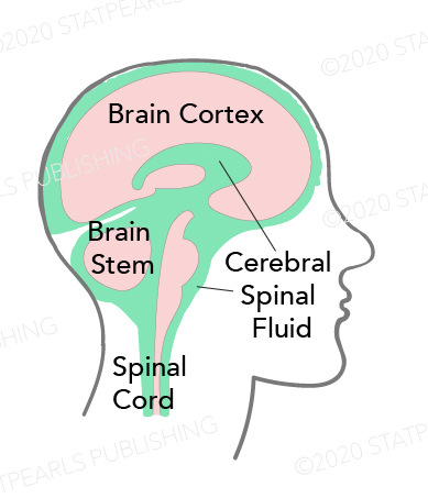
Cerebrospinal Fluid Distribution. The illustration shows cerebrospinal fluid in relation to the cerebral cortex, brainstem, and spinal cord.
Illustration by K Humphreys
(Click Image to Enlarge)
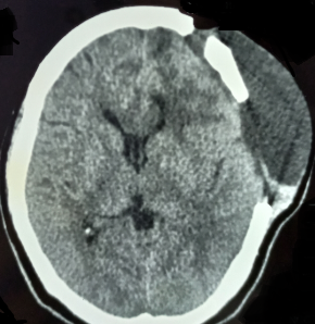
Pseudomeningocele, Computed Tomography. This image shows a cerebrospinal fluid leak, with fluid surrounding the brain and spinal cord.
Contributed by S Munakomi, MD
(Click Image to Enlarge)

Pseudomeningocele. This image shows a patient with a large pseudomeningocele.
Contributed by S Munakomi, MD
References
Theologou M, Natsis K, Kouskouras K, Chatzinikolaou F, Varoutis P, Skoulios N, Tsitouras V, Tsonidis C. Cerebrospinal Fluid Homeostasis and Hydrodynamics: A Review of Facts and Theories. European neurology. 2022:85(4):313-325. doi: 10.1159/000523709. Epub 2022 Apr 11 [PubMed PMID: 35405679]
Hutton D, Fadelalla MG, Kanodia AK, Hossain-Ibrahim K. Choroid plexus and CSF: an updated review. British journal of neurosurgery. 2022 Jun:36(3):307-315. doi: 10.1080/02688697.2021.1903390. Epub 2021 Apr 6 [PubMed PMID: 33821737]
Bulat M, Klarica M. Recent insights into a new hydrodynamics of the cerebrospinal fluid. Brain research reviews. 2011 Jan 1:65(2):99-112. doi: 10.1016/j.brainresrev.2010.08.002. Epub 2010 Sep 29 [PubMed PMID: 20817024]
Level 3 (low-level) evidenceYamada S. Cerebrospinal fluid dynamics. Croatian medical journal. 2021 Aug 31:62(4):399-410 [PubMed PMID: 34472743]
Brown PD, Davies SL, Speake T, Millar ID. Molecular mechanisms of cerebrospinal fluid production. Neuroscience. 2004:129(4):957-70 [PubMed PMID: 15561411]
Level 3 (low-level) evidenceMargetis K, Weisbrod LJ, Launico MV. Neuroanatomy, Cerebrospinal Fluid. StatPearls. 2025 Jan:(): [PubMed PMID: 29262203]
Dobrocky T, Nicholson P, Häni L, Mordasini P, Krings T, Brinjikji W, Cutsforth-Gregory JK, Schär R, Schankin C, Gralla J, Pereira VM, Raabe A, Farb R, Beck J, Piechowiak EI. Spontaneous intracranial hypotension: searching for the CSF leak. The Lancet. Neurology. 2022 Apr:21(4):369-380. doi: 10.1016/S1474-4422(21)00423-3. Epub 2022 Feb 25 [PubMed PMID: 35227413]
D'Antona L, Jaime Merchan MA, Vassiliou A, Watkins LD, Davagnanam I, Toma AK, Matharu MS. Clinical Presentation, Investigation Findings, and Treatment Outcomes of Spontaneous Intracranial Hypotension Syndrome: A Systematic Review and Meta-analysis. JAMA neurology. 2021 Mar 1:78(3):329-337. doi: 10.1001/jamaneurol.2020.4799. Epub [PubMed PMID: 33393980]
Level 1 (high-level) evidenceLe C, Strong EB, Luu Q. Management of Anterior Skull Base Cerebrospinal Fluid Leaks. Journal of neurological surgery. Part B, Skull base. 2016 Oct:77(5):404-11. doi: 10.1055/s-0036-1584229. Epub 2016 Jun 2 [PubMed PMID: 27648397]
Tang R, Mao S, Li D, Ye H, Zhang W. Treatment and Outcomes of Iatrogenic Cerebrospinal Fluid Leak Caused by Different Surgical Procedures. World neurosurgery. 2020 Nov:143():e667-e675. doi: 10.1016/j.wneu.2020.08.069. Epub 2020 Aug 15 [PubMed PMID: 32805467]
Scoffings DJ. Imaging of Acquired Skull Base Cerebrospinal Fluid Leaks. Neuroimaging clinics of North America. 2021 Nov:31(4):509-522. doi: 10.1016/j.nic.2021.05.009. Epub [PubMed PMID: 34689930]
Baugnon KL, Hudgins PA. Skull base fractures and their complications. Neuroimaging clinics of North America. 2014 Aug:24(3):439-65, vii-viii. doi: 10.1016/j.nic.2014.03.001. Epub 2014 May 24 [PubMed PMID: 25086806]
Banks CA, Palmer JN, Chiu AG, O'Malley BW Jr, Woodworth BA, Kennedy DW. Endoscopic closure of CSF rhinorrhea: 193 cases over 21 years. Otolaryngology--head and neck surgery : official journal of American Academy of Otolaryngology-Head and Neck Surgery. 2009 Jun:140(6):826-33. doi: 10.1016/j.otohns.2008.12.060. Epub 2009 Feb 28 [PubMed PMID: 19467398]
Level 2 (mid-level) evidenceBakhsh A, Elmolla M, Buxton N, Brodbelt A. Chronic CSF leak from lumbar-peritoneal shunt tract: A case report. Surgical neurology international. 2022:13():205. doi: 10.25259/SNI_1084_2021. Epub 2022 May 20 [PubMed PMID: 35673636]
Level 3 (low-level) evidenceHanna G, Pando A, Saela S, Emami AP. Cerebrospinal fluid (CSF) leak after elective lumbar spinal fusion: Who is at risk? European spine journal : official publication of the European Spine Society, the European Spinal Deformity Society, and the European Section of the Cervical Spine Research Society. 2022 Dec:31(12):3560-3565. doi: 10.1007/s00586-022-07383-9. Epub 2022 Sep 12 [PubMed PMID: 36094667]
Farnsworth PJ, Madhavan AA, Verdoorn JT, Shlapak DP, Johnson DR, Cutsforth-Gregory JK, Brinjikji W, Lehman VT. Spontaneous intracranial hypotension: updates from diagnosis to treatment. Neuroradiology. 2023 Feb:65(2):233-243. doi: 10.1007/s00234-022-03079-5. Epub 2022 Nov 7 [PubMed PMID: 36336758]
Kranz PG, Gray L, Malinzak MD, Amrhein TJ. Spontaneous Intracranial Hypotension: Pathogenesis, Diagnosis, and Treatment. Neuroimaging clinics of North America. 2019 Nov:29(4):581-594. doi: 10.1016/j.nic.2019.07.006. Epub 2019 Aug 26 [PubMed PMID: 31677732]
Luetzen N, Dovi-Akue P, Fung C, Beck J, Urbach H. Spontaneous intracranial hypotension: diagnostic and therapeutic workup. Neuroradiology. 2021 Nov:63(11):1765-1772. doi: 10.1007/s00234-021-02766-z. Epub 2021 Jul 23 [PubMed PMID: 34297176]
Palavani LB, Andrade CVF, Andrade RA, Alves E, Barros MF, Barbieri JF. Cerebrospinal fluid fistula as a complication of reverse transcriptase-polymerase chain reaction collection for the detection of coronavirus disease 2019: illustrative cases. Journal of neurosurgery. Case lessons. 2023 Jan 30:5(5):. pii: CASE22478. doi: 10.3171/CASE22478. Epub 2023 Jan 30 [PubMed PMID: 36718870]
Level 3 (low-level) evidenceXing QQ, Miao M, Zhang QW, Wu Y, He FF. Gorham-Stout disease affecting the spine with cerebrospinal fluid leakage and Chiari-like tonsillar herniation: a rare case report and review of literature. BMC neurology. 2023 Feb 3:23(1):59. doi: 10.1186/s12883-023-03092-y. Epub 2023 Feb 3 [PubMed PMID: 36737721]
Level 3 (low-level) evidenceSchievink WI, Maya MM, Jean-Pierre S, Nuño M, Prasad RS, Moser FG. A classification system of spontaneous spinal CSF leaks. Neurology. 2016 Aug 16:87(7):673-9. doi: 10.1212/WNL.0000000000002986. Epub 2016 Jul 20 [PubMed PMID: 27440149]
Schievink WI, Maya MM. Diffuse non-aneurysmal SAH in spontaneous intracranial hypotension: Sequela of ventral CSF leak? Cephalalgia : an international journal of headache. 2016 May:36(6):589-92. doi: 10.1177/0333102415604473. Epub 2015 Sep 7 [PubMed PMID: 26346560]
Martínez-Capoccioni G, Serramito-García R, Martín-Bailón M, García-Allut A, Martín-Martín C. Spontaneous cerebrospinal fluid leaks in the anterior skull base secondary to idiopathic intracranial hypertension. European archives of oto-rhino-laryngology : official journal of the European Federation of Oto-Rhino-Laryngological Societies (EUFOS) : affiliated with the German Society for Oto-Rhino-Laryngology - Head and Neck Surgery. 2017 May:274(5):2175-2181. doi: 10.1007/s00405-017-4455-5. Epub 2017 Feb 7 [PubMed PMID: 28175991]
Englhard AS, Volgger V, Leunig A, Meßmer CS, Ledderose GJ. Spontaneous nasal cerebrospinal fluid leaks: management of 24 patients over 11 years. European archives of oto-rhino-laryngology : official journal of the European Federation of Oto-Rhino-Laryngological Societies (EUFOS) : affiliated with the German Society for Oto-Rhino-Laryngology - Head and Neck Surgery. 2018 Oct:275(10):2487-2494. doi: 10.1007/s00405-018-5089-y. Epub 2018 Aug 14 [PubMed PMID: 30109406]
Pagani-Estévez GL, Cutsforth-Gregory JK, Morris JM, Mokri B, Piepgras DG, Mauck WD, Eldrige JS, Watson JC. Procedural predictors of epidural blood patch efficacy in spontaneous intracranial hypotension. Regional anesthesia and pain medicine. 2019 Jan 13:():. pii: rapm-2018-000021. doi: 10.1136/rapm-2018-000021. Epub 2019 Jan 13 [PubMed PMID: 30636714]
Ommaya AK, Di Chiro G, Baldwin M, Pennybacker JB. Non-traumatic cerebrospinal fluid rhinorrhoea. Journal of neurology, neurosurgery, and psychiatry. 1968 Jun:31(3):214-25 [PubMed PMID: 5303103]
Schievink WI. Spontaneous Intracranial Hypotension. The New England journal of medicine. 2021 Dec 2:385(23):2173-2178. doi: 10.1056/NEJMra2101561. Epub [PubMed PMID: 34874632]
Ramakrishnan N, Roy R, Singh S, Goyal S, Gupta DK, Chugh R. Approach to Management of Cerebrospinal Fluid Rhinorrhea: Institutional Based Protocol. Indian journal of otolaryngology and head and neck surgery : official publication of the Association of Otolaryngologists of India. 2022 Oct:74(Suppl 2):737-744. doi: 10.1007/s12070-019-01728-5. Epub 2019 Aug 17 [PubMed PMID: 36452775]
Mamlouk MD, Shen PY, Jun P, Sedrak MF. Spontaneous Spinal CSF Leaks Stratified by Age, Body Mass Index, and Spinal Level. AJNR. American journal of neuroradiology. 2022 Jul:43(7):1068-1072. doi: 10.3174/ajnr.A7548. Epub 2022 Jun 23 [PubMed PMID: 35738670]
Taylor CA, Bell JM, Breiding MJ, Xu L. Traumatic Brain Injury-Related Emergency Department Visits, Hospitalizations, and Deaths - United States, 2007 and 2013. Morbidity and mortality weekly report. Surveillance summaries (Washington, D.C. : 2002). 2017 Mar 17:66(9):1-16. doi: 10.15585/mmwr.ss6609a1. Epub 2017 Mar 17 [PubMed PMID: 28301451]
Phang SY, Whitehouse K, Lee L, Khalil H, McArdle P, Whitfield PC. Management of CSF leak in base of skull fractures in adults. British journal of neurosurgery. 2016 Dec:30(6):596-604 [PubMed PMID: 27666293]
Stopa BM, Leyva OA, Harper CN, Truman KA, Corrales CE, Smith TR, Gormley WB. Decreased Incidence of CSF Leaks after Skull Base Fractures in the 21st Century: An Institutional Report. Journal of neurological surgery. Part B, Skull base. 2022 Feb:83(1):59-65. doi: 10.1055/s-0040-1716689. Epub 2020 Oct 16 [PubMed PMID: 35155071]
Menon SK, Onyia CU. A short review on a complication of lumbar spine surgery: CSF leak. Clinical neurology and neurosurgery. 2015 Dec:139():248-51. doi: 10.1016/j.clineuro.2015.10.013. Epub 2015 Oct 23 [PubMed PMID: 26523872]
Wong AP, Shih P, Smith TR, Slimack NP, Dahdaleh NS, Aoun SG, El Ahmadieh TY, Smith ZA, Scheer JK, Koski TR, Liu JC, Fessler RG. Comparison of symptomatic cerebral spinal fluid leak between patients undergoing minimally invasive versus open lumbar foraminotomy, discectomy, or laminectomy. World neurosurgery. 2014 Mar-Apr:81(3-4):634-40. doi: 10.1016/j.wneu.2013.11.012. Epub 2013 Nov 13 [PubMed PMID: 24239738]
Level 2 (mid-level) evidenceGupta KK, Balai E, Darr A, Jolly K. Reconstruction and Cerebrospinal Fluid Leaks in Endoscopic Endonasal Approach for the Management of Clival Chordomas-A Systematic Review. Indian journal of otolaryngology and head and neck surgery : official publication of the Association of Otolaryngologists of India. 2022 Dec:74(Suppl 3):4807-4815. doi: 10.1007/s12070-022-03114-0. Epub 2022 Jul 7 [PubMed PMID: 36742692]
Level 1 (high-level) evidenceKranz PG, Gray L, Malinzak MD, Houk JL, Kim DK, Amrhein TJ. CSF-Venous Fistulas: Anatomy and Diagnostic Imaging. AJR. American journal of roentgenology. 2021 Dec:217(6):1418-1429. doi: 10.2214/AJR.21.26182. Epub 2021 Jun 30 [PubMed PMID: 34191547]
Roytman M, Salama G, Robbins MS, Chazen JL. CSF-Venous Fistula. Current pain and headache reports. 2021 Jan 21:25(1):5. doi: 10.1007/s11916-020-00921-4. Epub 2021 Jan 21 [PubMed PMID: 33475890]
Wang EW, Vandergrift WA 3rd, Schlosser RJ. Spontaneous CSF Leaks. Otolaryngologic clinics of North America. 2011 Aug:44(4):845-56, vii. doi: 10.1016/j.otc.2011.06.018. Epub [PubMed PMID: 21819875]
Mokri B, Aksamit AJ, Atkinson JL. Paradoxical postural headaches in cerebrospinal fluid leaks. Cephalalgia : an international journal of headache. 2004 Oct:24(10):883-7 [PubMed PMID: 15377320]
Level 3 (low-level) evidenceWicklund MR, Mokri B, Drubach DA, Boeve BF, Parisi JE, Josephs KA. Frontotemporal brain sagging syndrome: an SIH-like presentation mimicking FTD. Neurology. 2011 Apr 19:76(16):1377-82. doi: 10.1212/WNL.0b013e3182166e42. Epub [PubMed PMID: 21502595]
Level 2 (mid-level) evidenceFriedauer L, Rezny-Kasprzak B, Steinmetz H, du Mesnil de Rochemont R, Foerch C. Spinal dural leaks in patients with infratentorial superficial siderosis of the central nervous system-Refinement of a diagnostic algorithm. European journal of neurology. 2022 Apr:29(4):1136-1144. doi: 10.1111/ene.14611. Epub 2020 Dec 14 [PubMed PMID: 33098710]
Meco C, Oberascher G, Arrer E, Moser G, Albegger K. Beta-trace protein test: new guidelines for the reliable diagnosis of cerebrospinal fluid fistula. Otolaryngology--head and neck surgery : official journal of American Academy of Otolaryngology-Head and Neck Surgery. 2003 Nov:129(5):508-17 [PubMed PMID: 14595273]
Bachmann G, Achtelik R, Nekic M, Michel O. [Beta-trace protein in diagnosis of cerebrospinal fluid fistula]. HNO. 2000 Jul:48(7):496-500 [PubMed PMID: 10955226]
Chan DT, Poon WS, IP CP, Chiu PW, goh KY. How useful is glucose detection in diagnosing cerebrospinal fluid leak? The rational use of CT and Beta-2 transferrin assay in detection of cerebrospinal fluid fistula. Asian journal of surgery. 2004 Jan:27(1):39-42 [PubMed PMID: 14719513]
Hiremath SB, Gautam AA, Sasindran V, Therakathu J, Benjamin G. Cerebrospinal fluid rhinorrhea and otorrhea: A multimodality imaging approach. Diagnostic and interventional imaging. 2019 Jan:100(1):3-15. doi: 10.1016/j.diii.2018.05.003. Epub 2018 Jun 15 [PubMed PMID: 29910174]
Chiapparini L, Ciceri E, Nappini S, Castellani MR, Mea E, Bussone G, Leone M, Savoiardo M. Headache and intracranial hypotension: neuroradiological findings. Neurological sciences : official journal of the Italian Neurological Society and of the Italian Society of Clinical Neurophysiology. 2004 Oct:25 Suppl 3():S138-41 [PubMed PMID: 15549524]
Signorelli F, Caccavella VM, Giordano M, Ioannoni E, Caricato A, Polli FM, Olivi A, Montano N. A systematic review and meta-analysis of factors affecting the outcome of the epidural blood patching in spontaneous intracranial hypotension. Neurosurgical review. 2021 Dec:44(6):3079-3085. doi: 10.1007/s10143-021-01505-5. Epub 2021 Feb 21 [PubMed PMID: 33611638]
Level 1 (high-level) evidenceTai YC, Tai YS, Ou CH, Lui CC, Wang HK, Kuo HC, Hsu SP. Treatment, Outcome, and Relapse of Spontaneous and Nonspontaneous Cerebrospinal Fluid Leak. Brain sciences. 2022 Mar 2:12(3):. doi: 10.3390/brainsci12030340. Epub 2022 Mar 2 [PubMed PMID: 35326296]
Bernal-Sprekelsen M, Bleda-Vázquez C, Carrau RL. Ascending meningitis secondary to traumatic cerebrospinal fluid leaks. American journal of rhinology. 2000 Jul-Aug:14(4):257-9 [PubMed PMID: 10979500]
Level 2 (mid-level) evidencePatel EA, Saad AM, Filip P, Filimonov A. Acetazolamide use in postoperative CSF leak prevention: A literature review. American journal of otolaryngology. 2025 Jul-Aug:46(4):104656. doi: 10.1016/j.amjoto.2025.104656. Epub 2025 Apr 28 [PubMed PMID: 40311494]
Choi SY, Seong M, Kim EY, Youn MS, Cho S, Jang H, Lee MJ. Outcome of epidural blood patch for imaging-negative spontaneous intracranial hypotension. Cephalalgia : an international journal of headache. 2023 Feb:43(2):3331024221140471. doi: 10.1177/03331024221140471. Epub [PubMed PMID: 36739515]
Khan R, Sajjad M, Khan AA, Ahmad B, Ahmad S, Mushtaq M, Sultan S, Iftikhar O. Comparison Of Lumbar Drain Insertion And Conservative Management In The Treatment Of Traumatic CSF Rhinorrhoea. Journal of Ayub Medical College, Abbottabad : JAMC. 2019 Jul-Sep:31(3):441-444 [PubMed PMID: 31535524]
Cai X, Yang J, Zhu J, Tang C, Cong Z, Liu Y, Ma C. Reconstruction strategies for intraoperative CSF leak in endoscopic endonasal skull base surgery: systematic review and meta-analysis. British journal of neurosurgery. 2022 Aug:36(4):436-446. doi: 10.1080/02688697.2020.1849548. Epub 2021 Jan 21 [PubMed PMID: 33475004]
Level 1 (high-level) evidenceCRANIAL Consortium. CSF rhinorrhoea after endonasal intervention to the skull base (CRANIAL): A multicentre prospective observational study. Frontiers in oncology. 2022:12():1049627. doi: 10.3389/fonc.2022.1049627. Epub 2023 Jan 4 [PubMed PMID: 36688936]
Level 2 (mid-level) evidenceMirza S, Thaper A, McClelland L, Jones NS. Sinonasal cerebrospinal fluid leaks: management of 97 patients over 10 years. The Laryngoscope. 2005 Oct:115(10):1774-7 [PubMed PMID: 16222193]
Perez-Roman RJ, Bryant JP, Tapamo HJ, Luther E, Levene HB. Use of an External Ventricular Drain for Treatment of a Thoracolumbar Cerebrospinal Fluid Leak: A Case Report and Review of Literature. Cureus. 2022 Apr:14(4):e24066. doi: 10.7759/cureus.24066. Epub 2022 Apr 12 [PubMed PMID: 35573571]
Level 3 (low-level) evidencePlatt MP, Parnes SM. Management of unexpected cerebrospinal fluid leak during endoscopic sinus surgery. Current opinion in otolaryngology & head and neck surgery. 2009 Feb:17(1):28-32. doi: 10.1097/MOO.0b013e32831fb593. Epub [PubMed PMID: 19225302]
Level 3 (low-level) evidenceTorres-Bayona S, Velasquez N, Nakassa A, Eguiluz-Melendez A, Hernandez V, Vega B, Borghei-Razavi H, Miranda-Acosta Y, Wang EW, Snyderman CH, Gardner PA. Risk Factors and Reconstruction Techniques for Persistent Cerebrospinal Fluid Leak in Patients Undergoing Endoscopic Endonasal Approach to the Posterior Fossa. Journal of neurological surgery. Part B, Skull base. 2022 Jun:83(Suppl 2):e318-e323. doi: 10.1055/s-0041-1729904. Epub 2021 May 17 [PubMed PMID: 35832933]
Knight A. The differential diagnosis of rhinorrhea. The Journal of allergy and clinical immunology. 1995 May:95(5 Pt 2):1080-3 [PubMed PMID: 7751525]
Schievink WI. Spontaneous spinal cerebrospinal fluid leaks and intracranial hypotension. JAMA. 2006 May 17:295(19):2286-96 [PubMed PMID: 16705110]
Daele JJ, Goffart Y, Machiels S. Traumatic, iatrogenic, and spontaneous cerebrospinal fluid (CSF) leak: endoscopic repair. B-ENT. 2011:7 Suppl 17():47-60 [PubMed PMID: 22338375]
Charalambous LT, Rajkumar S, Liu B, Adil SM, Wong M, Hodges S, Amrhein TJ, Leithe LG, Parente B, Lee HJ, Lad SP. Treatment Patterns and Health Care Resource Utilization of Iatrogenic Spinal Cerebrospinal Fluid Leaks in the United States. Clinical spine surgery. 2022 Nov 1:35(9):E725-E730. doi: 10.1097/BSD.0000000000001363. Epub 2022 Jul 14 [PubMed PMID: 35858207]
Yadav YR, Parihar V, Janakiram N, Pande S, Bajaj J, Namdev H. Endoscopic management of cerebrospinal fluid rhinorrhea. Asian journal of neurosurgery. 2016 Jul-Sep:11(3):183-93. doi: 10.4103/1793-5482.145101. Epub [PubMed PMID: 27366243]
Soni AJ, Modi G. Outcome of uncorrected CSF leak and consequent recurrent meningitis in a patient: a case presentation and literature review. British journal of neurosurgery. 2020 Oct:34(5):492-494. doi: 10.1080/02688697.2018.1478063. Epub 2018 May 28 [PubMed PMID: 29807467]
Level 3 (low-level) evidenceDaudia A, Biswas D, Jones NS. Risk of meningitis with cerebrospinal fluid rhinorrhea. The Annals of otology, rhinology, and laryngology. 2007 Dec:116(12):902-5 [PubMed PMID: 18217509]