Introduction
Bowenoid papulosis is a premalignant skin condition transmitted through sexual contact and caused by oncogenic human papillomavirus (HPV) types. Lloyd first described the condition in 1970 as a pigmented Bowen disease of the groin in a young man. The lesions shared histologic features with Bowen disease but demonstrated distinct clinical characteristics.[1] In 1977, Kopf and Bart reported multiple recurrent papules showing epidermal hyperplasia and atypical keratinocytes throughout the epithelium. The findings were consistent with squamous cell carcinoma in situ (SCCIS), and previous photochemotherapy was hypothesized as a contributing factor.[2]
In 1978, Wade and colleagues examined genital lesions in young adults that resembled condylomas, lichen planus, or psoriasis. Histologic analysis revealed unequivocal features of SCCIS. The authors subsequently named the condition, previously described by Lloyd, Kopf, and Bart, as Bowenoid papulosis.[3]
Bowenoid papulosis is caused by high-risk HPV serotypes, particularly 16 and 18. Additional contributing factors such as tobacco use, hormonal fluctuations, and local trauma have also been proposed. The condition primarily affects young adults, with a higher prevalence among men.[4] Clinically, lesions appear as benign-appearing warty papules or plaques, occasionally violaceous in color, often mimicking lichen planus. Key differential diagnoses include lichen planus, condylomata acuminata, and psoriasis (see Image. Bowenoid Papulosis).
Diagnosis relies on clinical evaluation with confirmation by histologic examination. In contrast to Bowen disease, which frequently progresses to invasive squamous cell carcinoma (ISCC), Bowenoid papulosis rarely advances to invasive malignancy. Histologically, Bowenoid papulosis is now recognized as a high-grade squamous intraepithelial lesion (HSIL), typically exhibiting milder cytologic atypia than classic Bowen disease. Classification depends on anatomical location, such as the penis, vulva, or anal region.[5][6] Treatment options include topical chemotherapeutic agents, ablative modalities, and surgical excision. The biological behavior and long-term course of Bowenoid papulosis are uncertain. However, most lesions respond to therapy or undergo spontaneous regression.
Etiology
Register For Free And Read The Full Article
 Search engine and full access to all medical articles
- 10 free questions in your specialty
- Free CME/CE Activities
- Free daily question in your email
- Save favorite articles to your dashboard
- Emails offering discounts
Learn more about a Subscription to StatPearls Point-of-Care
Etiology
Bowenoid papulosis is a premalignant skin condition transmitted through sexual contact and caused by oncogenic types of HPV. Most lesions are associated with high-risk HPV types, particularly 16. Less frequently, HPV genotypes 18, 31 through 35, 39, 40, 42, 49, 51 through 54, and 56 have been identified.[7][8] The condition may also develop in immunocompromised individuals, including organ transplant recipients. Additional contributing factors include tobacco use, particularly in HPV-negative cases.[9][10] The relevance of poor hygiene, chronic maceration, and other local trauma is uncertain, and no single nonviral factor has been established as a definitive cause.
Epidemiology
Bowenoid papulosis commonly occurs among sexually active adults. Although rare, nonsexual transmission via fomites has been hypothesized in cases lacking sexual exposure and confirmed HPV infection. The incubation period ranges from several weeks to months and, in rare cases, years.[11] The condition slightly favors men over women, although no large cohort studies have established prevalence.
Onset typically occurs between 20 and 30 years of age, with a mean of 31 years, substantially younger than the average onset for Bowen disease, which more often affects men over 50 years.[12] Rare pediatric and geriatric cases have been documented, including a 3-year-old girl diagnosed in 1989 and a recent case involving a 63-year-old man.[13] Past reports noted higher occurrence in pregnant women, though incidence appears to have declined, likely due to widespread HPV vaccination among women of reproductive age.[14]
Many current diagnoses overlap histologically with HSIL of the penis, vulva, and anus. Among men who have sex with men (MSM), the estimated prevalence of anal HSIL is approximately 89 per 100,000, compared to 1 to 2 per 100,000 in the general population.[15][16] Penile HSIL affects an estimated 1 to 4 per 100,000 men.[17] Meanwhile, vulvar HSIL occurs in 3 to 5 per 100,000 women.[18] Insufficient epidemiologic data exist to define the global prevalence or demographic distribution of Bowenoid papulosis. As with other sexually transmitted infections, regional differences may reflect disparities in public health infrastructure.
Pathophysiology
The disease mechanism appears similar to the oncogenic process observed in other HPV-related conditions, including cervical cancer. Pathogenesis begins with viral entry and infection, followed by genomic alterations. Although progression to invasive carcinoma is rare in Bowenoid papulosis, viral oncoproteins E6 and E7 promote carcinogenesis by inactivating genome replication and cell cycle regulatory proteins such as p53 and Rb. The result is uncontrolled cell proliferation and impaired apoptosis.
HPV has been identified in both lesional and clinically normal skin in cases of Bowenoid papulosis and condylomata acuminata. This observation suggests that HPV infection alone may be insufficient to induce clinical disease. Additional host-related or environmental cofactors likely contribute to pathogenesis. The reduction in recurrence rates following smoking cessation supports this theory.[19] In a case series reported by Ranki et al, 6 of 14 patients with Bowenoid papulosis exhibited p53 mutations.[20]
Histopathology
Bowenoid papulosis is sometimes reported as histologically indistinguishable from Bowen disease. However, some authors believe the condition is a milder and more localized form of Bowen disease. The hallmark histologic pattern is full-thickness epidermal dysplasia, previously termed "Bowenoid dysplasia," characterized by apoptotic keratinocyte crowding and loss of polarity. Additional common findings include hyperkeratosis, psoriasiform hyperplasia, clubbing of the rete ridges, and parakeratosis. The basement membrane remains intact, consistent with SCCIS, as also observed in Bowen disease.[21]
At the cellular level, characteristic findings include hyperchromasia, rounded pyknotic nuclei, prominent nucleoli, metaphase mitotic figures, melanophages, multinucleated cells, and additional features consistent with chronic inflammation. Small, rounded, eosinophilic, inclusion-like bodies may be observed in the stratum corneum and granulosum, although true koilocytes are uncommon.[21][22] Follicular involvement is rare in Bowenoid papulosis, in contrast to Bowen disease.
Gross et al, in a 1985 publication, described 3 distinct growth patterns: flat (most common), papillary exophytic, and papillary endophytic. A distinguishing feature of Bowenoid papulosis is the alternation between areas of atypia and benign epithelial proliferation, particularly evident during spontaneous regression. Secondary localized cutaneous amyloidosis has been reported in biopsy specimens from Bowen disease. In several cases, patients had applied destructive topical substances, suggesting a potential link between this finding and apoptosis or necrosis induced by exogenous agents.[23]
Immunohistochemical staining may detect HPV-related nuclear antigens, sometimes seen on the surface of enucleated corneocytes. Expression of p16, a highly specific marker, may appear in both lesional and nonlesional skin in Bowenoid papulosis and condylomata acuminata.[24] Loss of S100 protein has been reported in both conditions and may be associated with neoplastic progression.[25][26] Electron microscopy reveals a reduced number of intact desmosomes.
History and Physical
The chief complaint during clinical consultations often concerns the recent onset and spread of lesions in sensitive areas, including the vulva, penis (glans and scrotum), perianal region, groin, and gluteal area. Mild pruritus or a burning sensation may accompany these lesions. Bowenoid papulosis typically presents with a benign appearance. The condition is characterized by multiple, confluent, and multicentric papules that are partially pigmented or skin-colored, usually measuring less than 1 cm in diameter. Lesions most commonly affect the anogenital region bilaterally.
Pigmentation varies according to skin phototype, appearing pink or slightly red in lighter phototypes and deeply pigmented in darker phototypes. Lesion surfaces may be slightly rough or verrucous, complicating clinical differentiation from condyloma acuminata (see Image. Verrucous Plaque in Bowenoid Papulosis). A velvety texture is frequently observed in the vulvar area. Deep pigmentation more often affects the external genitalia, including the clitoris and major and minor labia, whereas the vaginal introitus mucosa tends to display a more leukoplakic appearance. In certain cases, papules may be shiny and dome-shaped (see Image. Bowenoid Papulosis with Multifocal Involvement).
Uncommon sites of Bowenoid papulosis include the fingers, periungual skin, neck, elbows, abdomen, shoulder, chin, and oral cavity.[27][28][29] These atypical presentations may result from sexual contact, fomite transmission, or autoinoculation from other affected areas.
In a series of case reports, Kupetzky described oral Bowenoid papulosis as clinically similar to its anogenital counterpart but noted additional features such as red velvety papules, nodules, and leukoplakia.[30] Since these findings overlap with other forms of oral SCCIS, diagnosis in this region is particularly challenging. Rare presentations also include linear Bowenoid papulosis, possibly associated with the Koebner phenomenon.[31] In the anterior prepuce, Bowenoid papulosis may present with the “Xiuli” phenomenon, a distinctive circular distribution of lesions that spares the inner plate and glans. The Xiuli pattern is strongly associated with coexisting diabetes mellitus.[32]
Evaluation
Clinical evaluation is the cornerstone of diagnosis. However, the typically bland appearance of Bowenoid papulosis and its strong resemblance to benign dermatological conditions frequently necessitate biopsy for accurate confirmation. Histological features may mirror those of Bowen disease, but factors such as younger age of onset, genital localization, and viral infection in nonsun-exposed areas support a diagnosis of Bowenoid papulosis. Additional diagnostic methods are discussed below.
Virologic studies, including polymerase chain reaction and in situ hybridization, assist in distinguishing Bowenoid papulosis from its primary differential diagnosis, condyloma acuminatum. These methods enable identification of HPV subtypes, which differ between the 2 entities, HPV 6 and 11 in condyloma versus high-risk genotypes like HPV 16 in Bowenoid papulosis. Evaluation and ongoing monitoring of both male and female sexual partners, including screening for HPV 16, have been strongly recommended by multiple authors.[33]
Although not significantly increasing diagnostic accuracy, dermoscopy offers value in excluding serious pigmented lesions, such as mucosal melanoma and extramammary Paget disease. This diagnostic tool may also aid in differentiating from benign disorders like lichen planus, which display distinctive dermoscopic features. In 2021, Ürün et al reviewed the dermoscopic features of Bowenoid papulosis and concluded that no consistent pattern has been established due to the wide variation in clinical presentation. The most frequently reported findings include glomerular and dotted vascular structures, along with brown and gray dots showing linear distribution.[34]
Reflectance confocal microscopy, when available, offers potential for same-day diagnosis and follow-up in dermatology settings. Anoscopy should be performed in individuals with a history of receptive anal intercourse, regardless of the current location of Bowenoid papulosis lesions.
Treatment / Management
Treatment aims to prevent malignant transformation while minimizing the need for extensive tissue excision, which may result in anatomical or functional complications. No formal guidelines have been established for the management of Bowenoid papulosis, and no ablative modality has demonstrated superiority. Treatment approaches are explained below.
Locally destructive methods, including simple excision, electrocoagulation, cryosurgery, application of liquid nitrogen, podophyllin, and electrodesiccation, are among the most effective interventions. Carbon dioxide laser vaporization is generally discouraged in HPV-associated lesions due to the risk of aerosolizing viral particles and potential inhalation exposure.[35](A1)
Topical therapies such as 5-fluorouracil and 5% imiquimod offer self-administered alternatives. Shimizu et al reported complete lesion clearance following 2 months of 5% imiquimod therapy.[36] In another case described by Shaw et al, cryotherapy combined with imiquimod, applied 2 to 5 times weekly, followed by Mohs micrographic surgery, resulted in complete resolution after 12 months.[37](B3)
Photodynamic therapy remains underutilized but offers favorable cosmetic outcomes and short recovery periods.[38] Experimental treatments have included local injections of diluted aqueous bleomycin sulfate.(B2)
In pregnancy, when lesions involve the vulvar or perianal region, deferring treatment until postpartum and considering cesarean delivery may be appropriate.[39] Expectant management is often favored due to the potential for spontaneous regression during or after pregnancy.(B3)
Combination strategies—for example, integrating topical agents such as imiquimod with ablative methods like cryotherapy or surgical excision, or combining photodynamic therapy with laser modalities such as holmium laser—may improve overall clearance.[40] Major ablative surgeries are not routinely indicated. However, Mohs surgery has achieved success in rare, treatment-resistant cases. Given the potential for spontaneous regression, conservative management remains a viable option for selected individuals.
Differential Diagnosis
The differential diagnosis of Bowenoid papulosis encompasses numerous inflammatory, infectious, and neoplastic dermatoses affecting the anogenital region. Accurate differentiation relies on histopathological analysis and, when indicated, adjunctive diagnostic tools. The following conditions should be considered when evaluating lesions with similar clinical morphology:
- Condylomata acuminata (genital warts): Caused by low-risk HPV types 6 and 11. Clinically similar to Bowenoid papulosis but lacking dysplastic histology and oncogenic potential.
- SCCIS: Includes erythroplasia of Queyrat and Bowen disease. Shares histologic features with Bowenoid papulosis but differs in anatomic distribution and malignant potential.
- Lichen planus: Inflammatory mucocutaneous disease with histologic hallmarks such as wedge-shaped hypergranulosis and characteristic Wickham striae.[41]
- Plasmacytic (Zoon) balanitis: Moist, velvety erythematous plaques on the glans penis or sulcus. Histology reveals dense plasmacytic infiltrates.[42]
- Lichen sclerosus et atrophicus: Chronic inflammatory disorder presenting with atrophic, hypopigmented plaques, often involving the vulvar or perianal epithelium.[43]
- Seborrheic keratosis: Benign epidermal neoplasm common in older individuals. Resembles genital warts or Bowenoid papulosis but lacks cytologic atypia.[44]
- Psoriasis: Erythematous plaques in intertriginous regions, occasionally mistaken for Bowenoid papulosis when typical scales are absent.[45]
- Melanocytic nevus: Benign melanocytic proliferation occasionally mimicking pigmented variants of Bowenoid papulosis.[46]
- Warty dyskeratoma: Rare, solitary keratotic lesion with follicular involvement, often misdiagnosed as condyloma or fibroepithelial polyp.[47]
Many of these conditions exhibit considerable phenotypic overlap with Bowenoid papulosis, particularly in early or atypical presentations. Careful correlation of clinical, histologic, and virologic data is required to avoid diagnostic error.
Prognosis
The general prognosis of Bowenoid papulosis is favorable, though its biological behavior is not fully defined. Clinical course varies, with spontaneous regression reported in some cases, especially during pregnancy or the postpartum period, as mentioned.[48] Lesions can also persist for several years, particularly in immunocompromised individuals, and rarely progress to ISCC. Inadequately treated lesions may recur locally. Reported mean disease duration ranges from 2.4 to 3.6 years.
Complications
The risk of progression from Bowenoid papulosis to ISCC is very low, with most estimates below 1%. Some studies have reported a maximum malignant transformation rate of up to 2.6%. Female sexual partners of affected men face an elevated risk of cervical infection with oncogenic HPV, particularly HPV 16, and demonstrate an increased likelihood of developing cervical carcinoma. Persistent sexual exposure to an infected partner may hinder HPV clearance and potentially accelerate progression to cervical malignancy.
Deterrence and Patient Education
As with other sexually transmitted infections, consistent and proper condom use reduces the risk of HPV transmission in Bowenoid papulosis.[49] Baldwin et al have noted that strengthening the immune response through HPV vaccination is effective in preventing intraepithelial neoplasia of the anus, penis, and vulva. In some cases, vaccination may also facilitate lesion clearance, as observed in condylomata acuminata.[50] Sexual partners of individuals with Bowenoid papulosis should undergo regular screening and receive appropriate follow-up care.
Pearls and Other Issues
The management of intraepithelial neoplasias, including Bowenoid papulosis, erythroplasia of Queyrat, and Bowen disease, is generally more aggressive than that of benign condylomata. A targeted biopsy, such as a colposcopically directed sample, is always necessary before therapy to histologically exclude microinvasion.
In adults younger than 45 with Bowenoid papulosis or erythroplasia, superficial lesion eradication using cryotherapy or laser vaporization may be appropriate to avoid disfigurement. Among immunocompetent adults younger than 35, Bowenoid papulosis is often considered benign and self-limiting. However, prolonged monitoring is recommended to detect recurrences.
Most HPV vaccines contain virus-like particles of HPV-16 and HPV-18. These formulations protect against the 2 high-risk types responsible for approximately 70% of cervical malignancies and high-grade cervical intraepithelial neoplasia. Limited cross-protection may also extend to closely related types such as HPV types 31, 33, and 45.
The recently approved nonavalent vaccine includes virus-like particles for HPV types 6 and 11, as well as high-risk types 16, 18, 31, 33, 45, 52, and 58. These 7 high-risk types account for nearly 90% of cervical cancers. Despite vaccination, Pap smear screening remains essential due to the vaccine’s incomplete efficacy.[51]
Enhancing Healthcare Team Outcomes
Patient-centered care for individuals with Bowenoid papulosis requires coordinated involvement from dermatologists, gynecologists, urologists, proctologists, advanced practice providers, nurses, and the patient’s sexual partners. Clinicians must be equipped to diagnose, evaluate, and manage the condition using tools such as dermoscopy and, when available, reflectance confocal microscopy. Dermatologists provide expertise, particularly in selecting and administering topical or ablative therapies.
Ethical considerations play a central role in treatment selection, particularly given the condition's involvement of anatomically sensitive areas where aggressive therapy may compromise quality of life and sexual health. Respect for patient autonomy must guide decision-making. Each team member must have clearly defined responsibilities, contributing specialized expertise to optimize care.
Effective interprofessional communication fosters a collaborative environment in which concerns, questions, and clinical information are openly exchanged. Coordinated care is essential to ensure safe, efficient management. Physicians, advanced practitioners, nurses, and pharmacists must work in concert from diagnosis through follow-up. This collaboration minimizes delays, reduces clinical errors, and promotes outcomes that reflect patient-defined goals.
Media
(Click Image to Enlarge)
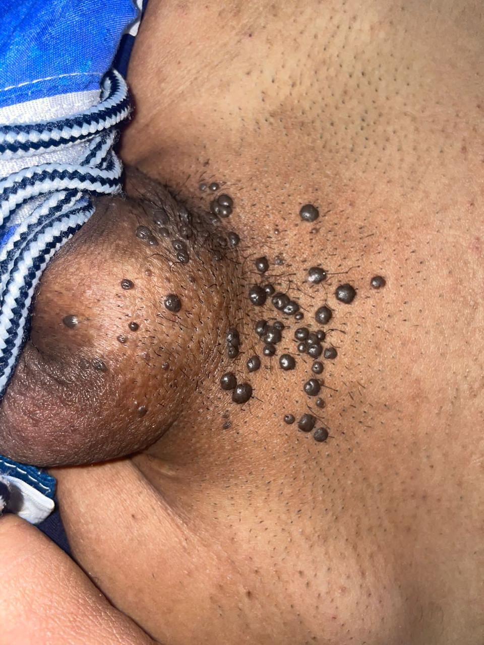
Bowenoid Papulosis. Multiple shiny, confluent brown papules are present on the pelvis and base of the penis.
Contributed by Tatiana Camayo, MD
(Click Image to Enlarge)
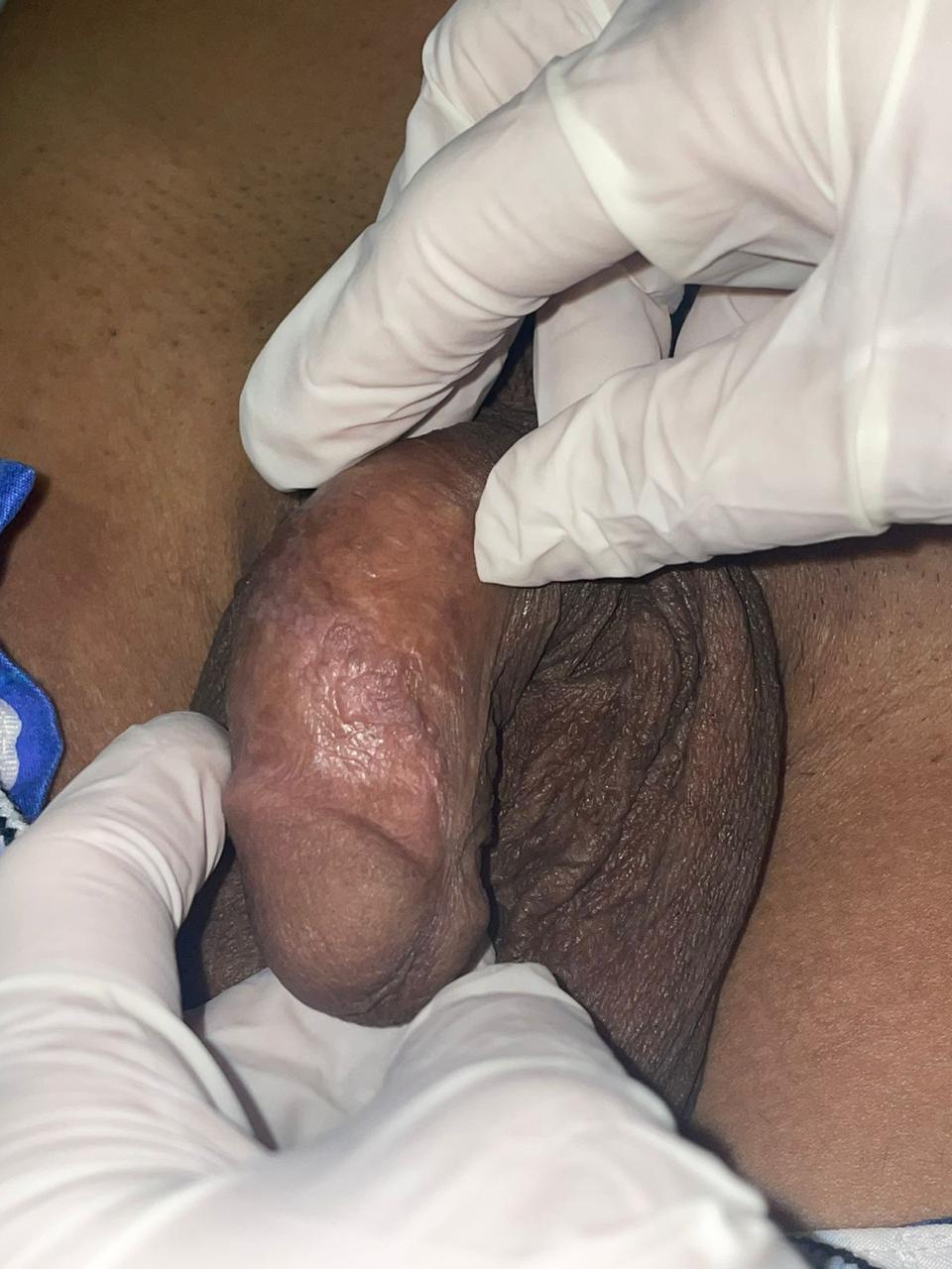
Verrucous Plaque in Bowenoid Papulosis. Multiple flat verrucous papules coalesce into a plaque on the penile shaft and balano-preputial sulcus, with histological confirmation of Bowenoid papulosis.
Contributed by Tatiana Camayo, MD
(Click Image to Enlarge)
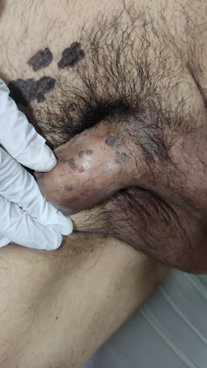
Bowenoid Papulosis with Multifocal Involvement. Multiple flat, brown, confluent verrucous papules are present on the penile shaft, with darker brown verrucous plaques extending onto the right inner thigh.
Contributed by Tatiana Camayo, MD
References
Lloyd KM. Multicentric pigmented Bowen's disease of the groin. Archives of dermatology. 1970 Jan:101(1):48-51 [PubMed PMID: 5416792]
Kopf AW, Bart RS. Tumor conference No. 11: multiple bowenoid papules of the penis: a new entity? The Journal of dermatologic surgery and oncology. 1977 May-Jun:3(3):265-9 [PubMed PMID: 874134]
Wade TR, Kopf AW, Ackerman AB. Bowenoid papulosis of the penis. Cancer. 1978 Oct:42(4):1890-903 [PubMed PMID: 361215]
Fader DJ, Stoler MH, Anderson TF. Isolated extragenital HPV-thirties-group-positive bowenoid papulosis in an AIDS patient. The British journal of dermatology. 1994 Oct:131(4):577-80 [PubMed PMID: 7947214]
Moch H, Cubilla AL, Humphrey PA, Reuter VE, Ulbright TM. The 2016 WHO Classification of Tumours of the Urinary System and Male Genital Organs-Part A: Renal, Penile, and Testicular Tumours. European urology. 2016 Jul:70(1):93-105. doi: 10.1016/j.eururo.2016.02.029. Epub 2016 Feb 28 [PubMed PMID: 26935559]
Stamm AW, Kobashi KC, Stefanovic KB. Urologic Dermatology: a Review. Current urology reports. 2017 Aug:18(8):62. doi: 10.1007/s11934-017-0712-9. Epub [PubMed PMID: 28667573]
Kato M, Shimizu A, Takeuchi Y, Hattori T, Abe M, Amano H, Motegi S, Tamura A, Ishikawa O. Human papillomaviruses in anogenital epithelial lesions. Acta dermato-venereologica. 2014 Sep:94(5):597-9. doi: 10.2340/00015555-1784. Epub [PubMed PMID: 24448688]
Go U, Miyata K, Nishimura-Yagi M, Mitsuishi T. Human papillomavirus 34 associated with bowenoid papulosis of the penile shaft. The Journal of dermatology. 2019 Jun:46(6):e192-e193. doi: 10.1111/1346-8138.14730. Epub 2018 Dec 20 [PubMed PMID: 30570779]
Dubina M, Goldenberg G. Viral-associated nonmelanoma skin cancers: a review. The American Journal of dermatopathology. 2009 Aug:31(6):561-73. doi: 10.1097/DAD.0b013e3181a58234. Epub [PubMed PMID: 19590418]
Vedhanayagam M, Rajagopalan R, Revathi K, Balamurugan BR, Srinivasahan KG, Damodaran D. Chronic extensive warty eruptions on the genital, pubic, crural, and perianal region. Indian journal of sexually transmitted diseases and AIDS. 2023 Jul-Dec:44(2):193-194. doi: 10.4103/ijstd.ijstd_103_23. Epub 2023 Dec 6 [PubMed PMID: 38223153]
Anic GM, Giuliano AR. Genital HPV infection and related lesions in men. Preventive medicine. 2011 Oct:53 Suppl 1(Suppl 1):S36-41. doi: 10.1016/j.ypmed.2011.08.002. Epub [PubMed PMID: 21962470]
Gross G, Hagedorn M, Ikenberg H, Rufli T, Dahlet C, Grosshans E, Gissmann L. Bowenoid papulosis. Presence of human papillomavirus (HPV) structural antigens and of HPV 16-related DNA sequences. Archives of dermatology. 1985 Jul:121(7):858-63 [PubMed PMID: 2990353]
Kazemi R, Jandaghi F, Derakhshan M, Bighamian M, Montazeri F. Bowenoid Papulosis of the Genitalia in an Older Individual: A Case Report. Advanced biomedical research. 2025:14():27. doi: 10.4103/abr.abr_286_24. Epub 2025 Mar 28 [PubMed PMID: 40303623]
Level 3 (low-level) evidenceBleeker MC, Heideman DA, Snijders PJ, Horenblas S, Dillner J, Meijer CJ. Penile cancer: epidemiology, pathogenesis and prevention. World journal of urology. 2009 Apr:27(2):141-50. doi: 10.1007/s00345-008-0302-z. Epub 2008 Jul 8 [PubMed PMID: 18607597]
Thitipatarakorn S, Teeratakulpisarn N, Nonenoy S, Klinsukontakul A, Suriwong S, Makphol J, Hongchookiat P, Chaya-Ananchot T, Chinlaertworasiri N, Mingkwanrungruang P, Sacdalan C, Poltavee K, Pankam T, Kerr SJ, Ramautarsing R, Colby D, Phanuphak N. Prevalence and incidence of anal high-grade squamous intraepithelial lesions in a cohort of cisgender men and transgender women who have sex with men diagnosed and treated during acute HIV acquisition in Bangkok, Thailand. Journal of the International AIDS Society. 2024 May:27(5):e26242. doi: 10.1002/jia2.26242. Epub [PubMed PMID: 38695517]
Palefsky JM, Lee JY, Jay N, Goldstone SE, Darragh TM, Dunlevy HA, Rosa-Cunha I, Arons A, Pugliese JC, Vena D, Sparano JA, Wilkin TJ, Bucher G, Stier EA, Tirado Gomez M, Flowers L, Barroso LF, Mitsuyasu RT, Lensing SY, Logan J, Aboulafia DM, Schouten JT, de la Ossa J, Levine R, Korman JD, Hagensee M, Atkinson TM, Einstein MH, Cracchiolo BM, Wiley D, Ellsworth GB, Brickman C, Berry-Lawhorn JM, ANCHOR Investigators Group. Treatment of Anal High-Grade Squamous Intraepithelial Lesions to Prevent Anal Cancer. The New England journal of medicine. 2022 Jun 16:386(24):2273-2282. doi: 10.1056/NEJMoa2201048. Epub [PubMed PMID: 35704479]
Soskin A, Vieillefond A, Carlotti A, Plantier F, Chaux A, Ayala G, Velazquez EF, Cubilla AL. Warty/basaloid penile intraepithelial neoplasia is more prevalent than differentiated penile intraepithelial neoplasia in nonendemic regions for penile cancer when compared with endemic areas: a comparative study between pathologic series from Paris and Paraguay. Human pathology. 2012 Feb:43(2):190-6. doi: 10.1016/j.humpath.2011.04.014. Epub 2011 Aug 10 [PubMed PMID: 21835427]
Level 2 (mid-level) evidencePreti M, Lewis F, Carcopino X, Bevilacqua F, Ellis LB, Halonen P, Hemida R, Jach R, Kesic V, Kyrgiou M, Maggino T, Pedro A, Querleu D, Stockdale C, Taumberger N, Temiz BE, Vieira-Baptista P, Gultekin M. Vulvar inspection at the time of cervical cancer screening: European Society of Gynaecological Oncology (ESGO), International Society for the Study of Vulvovaginal Disease (ISSVD), European College for the Study of Vulval Disease (ECSVD), and European Federation for Colposcopy (EFC) consensus statements. International journal of gynecological cancer : official journal of the International Gynecological Cancer Society. 2025 Jan:35(1):100007. doi: 10.1016/j.ijgc.2024.100007. Epub 2024 Dec 18 [PubMed PMID: 39878267]
Level 2 (mid-level) evidenceCoker AL, Bond SM, Williams A, Gerasimova T, Pirisi L. Active and passive smoking, high-risk human papillomaviruses and cervical neoplasia. Cancer detection and prevention. 2002:26(2):121-8 [PubMed PMID: 12102146]
Ranki A, Lassus J, Niemi KM. Relation of p53 tumor suppressor protein expression to human papillomavirus (HPV) DNA and to cellular atypia in male genital warts and in premalignant lesions. Acta dermato-venereologica. 1995 May:75(3):180-6 [PubMed PMID: 7653176]
Kimura S. Bowenoid papulosis of the genitalia. International journal of dermatology. 1982 Oct:21(8):432-6 [PubMed PMID: 6293984]
Cozma EC, Celarel AM, Stoenica IV, Lupu M, Banciu LM, Voiculescu VM. Correlations between Histopathological and Confocal Reflectance Microscopy Aspects in a Patient with Bowenoid Papulosis. Diagnostics (Basel, Switzerland). 2023 Apr 24:13(9):. doi: 10.3390/diagnostics13091531. Epub 2023 Apr 24 [PubMed PMID: 37174923]
Baykal C, Hurdogan O, Kobaner GB, Ekinci AP, Buyukbabani N. Secondary Localized Cutaneous Amyloidosis is not Rare in Bowen's Disease and Bowenoid Papulosis. Turk patoloji dergisi. 2022:38(1):54-59. doi: 10.5146/tjpath.2021.01530. Epub [PubMed PMID: 34514563]
Kazlouskaya V, Shustef E, Allam SH, Lal K, Elston D. Expression of p16 protein in lesional and perilesional condyloma acuminata and bowenoid papulosis: clinical significance and diagnostic implications. Journal of the American Academy of Dermatology. 2013 Sep:69(3):444-9. doi: 10.1016/j.jaad.2013.04.036. Epub 2013 May 21 [PubMed PMID: 23706650]
Elston DM. What is your diagnosis? Bowenoid papulosis. Cutis. 2010 Dec:86(6):278, 295-6 [PubMed PMID: 21284277]
Friedrich EG Jr. Reversible vulvar atypia. A case report. Obstetrics and gynecology. 1972 Feb:39(2):173-81 [PubMed PMID: 4333392]
Level 3 (low-level) evidenceMeira SF, Oncins R, Padgett E, Gracia-Cazaña T. Uncommon Location of Bowenoid Papulosis. Indian journal of dermatology. 2022 Nov-Dec:67(6):768-769. doi: 10.4103/ijd.ijd_527_21. Epub [PubMed PMID: 36998851]
McGrae JD Jr, Greer CE, Manos MM. Multiple Bowen's disease of the fingers associated with human papilloma virus type 16. International journal of dermatology. 1993 Feb:32(2):104-7 [PubMed PMID: 8382665]
Daley T, Birek C, Wysocki GP. Oral bowenoid lesions: differential diagnosis and pathogenetic insights. Oral surgery, oral medicine, oral pathology, oral radiology, and endodontics. 2000 Oct:90(4):466-73 [PubMed PMID: 11027384]
Kupetsky EA, Charles CA, Mones J. High-grade squamous intraepithelial lesion of the oral commissure (bowenoid papulosis). A case and review. Dermatology practical & conceptual. 2015 Oct:5(4):39-42. doi: 10.5826/dpc.0504a10. Epub 2015 Oct 31 [PubMed PMID: 26693089]
Level 3 (low-level) evidenceSweidan NA, Salman SM, Zaynoun ST, Sanaknaki BA, Kibbi AG. Linear bowenoid papulosis of the genitalia. A possible Koebner phenomenon. International journal of dermatology. 1990 Jul-Aug:29(6):430-1 [PubMed PMID: 2397968]
Huang J, Wang B, Shi L, Wang H. Clinical features of bowenoid papulosis of prepuce associated with diabetes mellitus. Photodiagnosis and photodynamic therapy. 2023 Jun:42():103536. doi: 10.1016/j.pdpdt.2023.103536. Epub 2023 Mar 24 [PubMed PMID: 36965760]
Henquet CJ. Anogenital malignancies and pre-malignancies. Journal of the European Academy of Dermatology and Venereology : JEADV. 2011 Aug:25(8):885-95. doi: 10.1111/j.1468-3083.2010.03969.x. Epub 2011 Jan 28 [PubMed PMID: 21272092]
Ürün YG, Ürün M, Fıçıcıoğlu S. A case of perianal bowenoid papulosis: dermoscopic features and a review of previous cases. Acta dermatovenerologica Alpina, Pannonica, et Adriatica. 2021 Mar:30(1):39-41 [PubMed PMID: 33765757]
Level 3 (low-level) evidencePalma S, Gnambs T, Crevenna R, Jordakieva G. Airborne human papillomavirus (HPV) transmission risk during ablation procedures: A systematic review and meta-analysis. Environmental research. 2021 Jan:192():110437. doi: 10.1016/j.envres.2020.110437. Epub 2020 Nov 9 [PubMed PMID: 33181134]
Level 1 (high-level) evidenceShimizu A, Kato M, Ishikawa O. Bowenoid papulosis successfully treated with imiquimod 5% cream. The Journal of dermatology. 2014 Jun:41(6):545-6. doi: 10.1111/1346-8138.12510. Epub [PubMed PMID: 24909216]
Shaw KS, Nguyen GH, Lacouture M, Deng L. Combination of imiquimod with cryotherapy in the treatment of penile intraepithelial neoplasia. JAAD case reports. 2017 Nov:3(6):546-549. doi: 10.1016/j.jdcr.2017.07.018. Epub 2017 Nov 6 [PubMed PMID: 29264388]
Level 3 (low-level) evidenceChe Q, Huang X, Li C, Li J, Jiang L, Zeng K. Effectiveness of photodynamic therapy with 5-aminolevulinic acid for Bowenoid papulosis: A retrospective study with long-term follow-up. Photodiagnosis and photodynamic therapy. 2022 Sep:39():102918. doi: 10.1016/j.pdpdt.2022.102918. Epub 2022 May 24 [PubMed PMID: 35618257]
Level 2 (mid-level) evidenceShastry V, Betkerur J, Kushalappa. Bowenoid papulosis of the genitalia successfully treated with topical tazarotene: a report of two cases. Indian journal of dermatology. 2009 Jul:54(3):283-6. doi: 10.4103/0019-5154.55643. Epub [PubMed PMID: 20161865]
Level 3 (low-level) evidenceGao J, Zhang H, Zhou Z, Sun X, Zhang G, Wang P, Wang X. The combination of holmium laser and ALA-PDT for Bowenoid Papulosis with diffuse large B-cell lymphoma. Photodiagnosis and photodynamic therapy. 2024 Feb:45():103953. doi: 10.1016/j.pdpdt.2023.103953. Epub 2023 Dec 23 [PubMed PMID: 38145769]
Kusari A, Ahluwalia J. Lichen Planus. The New England journal of medicine. 2018 Aug 9:379(6):567. doi: 10.1056/NEJMicm1802078. Epub [PubMed PMID: 30089065]
Relhan V, Kumar A, Kaur A. Zoon's Balanitis - Update of Clinical Spectrum and Management. Indian journal of dermatology. 2024 Jan-Feb:69(1):63-73. doi: 10.4103/ijd.ijd_834_22. Epub 2024 Feb 27 [PubMed PMID: 38572053]
Kraus CN. Vulvar Lichen Sclerosus. JAMA dermatology. 2022 Sep 1:158(9):1088. doi: 10.1001/jamadermatol.2022.0359. Epub [PubMed PMID: 35793083]
Barthelmann S, Butsch F, Lang BM, Stege H, Großmann B, Schepler H, Grabbe S. Seborrheic keratosis. Journal der Deutschen Dermatologischen Gesellschaft = Journal of the German Society of Dermatology : JDDG. 2023 Mar:21(3):265-277. doi: 10.1111/ddg.14984. Epub 2023 Mar 9 [PubMed PMID: 36892019]
Boehncke WH, Schön MP. Psoriasis. Lancet (London, England). 2015 Sep 5:386(9997):983-94. doi: 10.1016/S0140-6736(14)61909-7. Epub 2015 May 27 [PubMed PMID: 26025581]
Hauschild A, Egberts F, Garbe C, Bauer J, Grabbe S, Hamm H, Kerl H, Reusch M, Rompel R, Schlaeger M, expert group "Melanocytic nevi". Melanocytic nevi. Journal der Deutschen Dermatologischen Gesellschaft = Journal of the German Society of Dermatology : JDDG. 2011 Sep:9(9):723-34. doi: 10.1111/j.1610-0387.2011.07741.x. Epub 2011 Jul 15 [PubMed PMID: 21762380]
Lencastre A, Campos S, Cabete J. Warty dyskeratoma. Journal of the American Academy of Dermatology. 2016 Sep:75(3):e97-e98. doi: 10.1016/j.jaad.2016.02.1157. Epub [PubMed PMID: 27543244]
Skinner MS, Sternberg WH, Ichinose H, Collins J. Spontaneous regression of Bowenoid atypia of the vulva. Obstetrics and gynecology. 1973 Jul:42(1):40-6 [PubMed PMID: 4352642]
Hariri S, Warner L. Condom use and human papillomavirus in men. The Journal of infectious diseases. 2013 Aug 1:208(3):367-9. doi: 10.1093/infdis/jit193. Epub 2013 May 3 [PubMed PMID: 23644282]
Baldwin PJ, van der Burg SH, Boswell CM, Offringa R, Hickling JK, Dobson J, Roberts JS, Latimer JA, Moseley RP, Coleman N, Stanley MA, Sterling JC. Vaccinia-expressed human papillomavirus 16 and 18 e6 and e7 as a therapeutic vaccination for vulval and vaginal intraepithelial neoplasia. Clinical cancer research : an official journal of the American Association for Cancer Research. 2003 Nov 1:9(14):5205-13 [PubMed PMID: 14614000]
Joura EA, Giuliano AR, Iversen OE, Bouchard C, Mao C, Mehlsen J, Moreira ED Jr, Ngan Y, Petersen LK, Lazcano-Ponce E, Pitisuttithum P, Restrepo JA, Stuart G, Woelber L, Yang YC, Cuzick J, Garland SM, Huh W, Kjaer SK, Bautista OM, Chan IS, Chen J, Gesser R, Moeller E, Ritter M, Vuocolo S, Luxembourg A, Broad Spectrum HPV Vaccine Study. A 9-valent HPV vaccine against infection and intraepithelial neoplasia in women. The New England journal of medicine. 2015 Feb 19:372(8):711-23. doi: 10.1056/NEJMoa1405044. Epub [PubMed PMID: 25693011]